Experiencing a burst hemorrhoid without accompanying pain can be both confusing and concerning for patients. While most thrombosed external hemorrhoids cause intense discomfort before rupturing, some cases present with minimal or absent pain sensations, creating uncertainty about the appropriate response. This phenomenon occurs when the natural fibrinolytic processes within the hemorrhoidal tissue break down blood clots efficiently, allowing for spontaneous decompression without significant nociceptor activation.
The absence of pain during hemorrhoidal rupture doesn’t indicate a less serious condition, but rather suggests that the body’s natural healing mechanisms are functioning optimally. Understanding the underlying pathophysiology, recognising the clinical signs, and implementing appropriate management strategies are essential for ensuring proper healing and preventing complications. Modern proctological approaches emphasise conservative management for painless ruptures, focusing on tissue preservation and optimal wound healing conditions.
Understanding painless thrombosed external haemorrhoid rupture pathophysiology
The mechanism behind painless hemorrhoidal rupture involves complex vascular and neurological processes that differ significantly from typical thrombosed hemorrhoids. When external hemorrhoids develop blood clots, the resulting tissue distension typically activates pain receptors in the highly innervated perianal region. However, certain conditions can prevent this normal pain response, including rapid clot dissolution, adequate tissue oxygenation, and minimal inflammatory cascade activation.
Vascular anatomy of anal cushions and haemorrhoidal plexus
The hemorrhoidal plexus consists of arteriovenous communications that form cushioned structures supporting continence mechanisms. These vascular networks include the superior, middle, and inferior hemorrhoidal vessels, each with distinct drainage patterns and pressure characteristics. External hemorrhoids arise from the inferior hemorrhoidal plexus, positioned below the dentate line where somatic innervation provides acute pain sensation under normal circumstances.
Understanding this anatomy explains why some ruptures occur without pain—when venous drainage remains partially patent during thrombosis, tissue pressure doesn’t reach critical levels that activate nociceptors. The rich collateral circulation in the perianal region can maintain adequate perfusion, preventing the severe ischemia that typically causes intense pain in thrombosed hemorrhoids.
Thrombosis formation mechanisms in external haemorrhoidal vessels
Thrombosis formation in external hemorrhoids follows classical coagulation cascades, initiated by factors such as venous stasis, endothelial damage, and hypercoagulable states. Virchow’s triad perfectly describes the conditions leading to hemorrhoidal thrombosis: blood flow changes during straining, vessel wall trauma from chronic irritation, and altered blood composition due to dehydration or inflammatory states.
The clot formation process typically begins with platelet aggregation at sites of endothelial dysfunction, followed by fibrin deposition and red blood cell entrapment. However, in cases destined for painless rupture, the thrombotic process often remains localised, with surrounding tissues maintaining their normal architecture and innervation patterns, thus preserving comfort levels.
Natural fibrinolytic processes leading to spontaneous rupture
The body’s fibrinolytic system plays a crucial role in painless hemorrhoidal rupture through the activation of plasminogen to plasmin, which breaks down fibrin clots systematically. This natural process occurs more efficiently in some individuals due to genetic variations in fibrinolytic enzyme production, dietary factors influencing coagulation balance, or medications affecting the clotting cascade.
Spontaneous clot dissolution often occurs within 48-72 hours of thrombosis formation, creating a gradual pressure release that minimises tissue trauma and pain receptor activation.
Absence of nociceptor activation in Post-Rupture haemorrhoids
Pain absence during hemorrhoidal rupture indicates that nociceptors—specialised nerve endings that detect harmful stimuli—remain largely unactivated throughout the process. This can occur when tissue pressure increases gradually rather than acutely, allowing nerve adaptation, or when anti-inflammatory mediators counteract typical pain-inducing substances like prostaglandins and cytokines.
The perianal region contains various nociceptor types, including mechanoreceptors sensitive to pressure changes and chemoreceptors responding to inflammatory mediators. When rupture occurs through natural fibrinolytic processes rather than sudden pressure buildup, these receptors may not reach their activation thresholds, resulting in the notably painless presentation that patients experience.
Clinical assessment of ruptured haemorrhoidal tissue without pain sensation
Proper clinical assessment of painless ruptured hemorrhoids requires systematic evaluation to differentiate this condition from other perianal pathologies. The absence of pain doesn’t eliminate the need for thorough examination, as complications can develop silently, and other serious conditions may present similarly. Healthcare providers must maintain heightened awareness when patients report bleeding without discomfort, as this combination can indicate various underlying conditions.
Visual examination techniques for perianal haematoma resolution
Visual inspection remains the cornerstone of hemorrhoidal assessment, particularly for external hemorrhoids that have ruptured. The examination should be conducted in good lighting with the patient in a comfortable position, typically left lateral or knee-chest position. Look for signs of recent rupture including irregular tissue edges, residual hematoma, and the characteristic bluish-purple discoloration that gradually resolves.
Post-rupture hemorrhoids often present with a deflated appearance, where previously tense, swollen tissue now appears flaccid with possible surface irregularities. The surrounding skin may show signs of stretching or minor surface breaks where the clot evacuated. Document the size, location, and appearance of any residual swelling, as this information guides ongoing management decisions.
Differentiation from anal fissures and perirectal abscess drainage
Distinguishing ruptured hemorrhoids from anal fissures requires careful attention to location, appearance, and associated symptoms. Anal fissures typically occur at the posterior or anterior midline, appearing as linear tears with well-defined edges, while ruptured hemorrhoids present as irregular openings in previously swollen tissue masses. The bleeding pattern also differs—fissures cause sharp pain during defecation, whereas painless hemorrhoid rupture may produce intermittent bleeding.
Perirectal abscess drainage can mimic hemorrhoid rupture, particularly when pain is minimal due to effective decompression. However, abscess drainage typically produces purulent material rather than pure blood, and patients often have a history of fever or systemic symptoms. The presence of granulation tissue and ongoing purulent discharge suggests abscess drainage rather than simple hemorrhoid rupture.
Assessing residual thrombotic material and tissue viability
Evaluation of residual thrombotic material requires gentle palpation to identify any remaining firm clots within the hemorrhoidal tissue. Viable tissue maintains its normal pink coloration and soft consistency, while necrotic areas appear darker and may feel firmer or more friable. The presence of extensive necrotic tissue may require surgical debridement to prevent infection and promote healing.
Tissue viability assessment also involves checking capillary refill in the surrounding area and noting any signs of compromised circulation. Healthy tissue should demonstrate good perfusion with prompt capillary refill and absence of progressive discoloration. Monitor for signs of secondary infection, including increased warmth, spreading erythema, or development of purulent discharge.
Digital rectal examination considerations Post-Rupture
Digital rectal examination following hemorrhoid rupture should be approached cautiously, particularly when tissue integrity may be compromised. The examination helps assess internal hemorrhoids, rectal tone, and rule out other pathology, but must be performed gently to avoid further tissue trauma. Use adequate lubrication and proceed slowly, stopping if patient discomfort develops or tissue resistance is encountered.
During the examination, palpate for any internal masses, assess sphincter function, and check for tenderness or irregularities in the rectal wall. The absence of pain doesn’t guarantee tissue stability, so maintain gentle pressure throughout the examination. Document any findings including sphincter tone, presence of internal hemorrhoids, and overall tissue condition.
Immediate conservative management protocols for burst haemorrhoids
Conservative management represents the gold standard for treating painless ruptured hemorrhoids, focusing on creating optimal conditions for natural healing while preventing complications. The approach emphasises gentle cleansing, maintaining tissue hydration, and supporting the body’s natural repair mechanisms. Most patients respond well to conservative measures, avoiding the need for surgical intervention while achieving excellent functional outcomes.
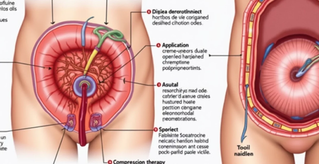
Topical haemostatic agents and antiseptic cleansing solutions
Application of appropriate haemostatic agents can help control bleeding and promote clot formation at the rupture site. Phenylephrine-based preparations provide dual benefits through vasoconstriction and mild anaesthetic properties, though the latter may be less relevant in painless cases. Witch hazel solutions offer natural astringent properties that help reduce tissue inflammation and provide gentle antiseptic action.
Cleansing protocols should emphasise gentle antiseptic solutions that don’t compromise tissue healing. Dilute povidone-iodine solutions or chlorhexidine preparations can be used sparingly, but avoid alcohol-based products that may cause tissue desiccation. The key principle involves maintaining cleanliness without disrupting the natural healing environment or causing additional tissue trauma.
Sitz bath temperature regulation and frequency guidelines
Sitz baths represent a cornerstone of hemorrhoidal care, promoting circulation, reducing inflammation, and maintaining optimal hygiene around the affected area. Water temperature should be maintained between 37-40°C (98-104°F) to ensure therapeutic benefit without causing tissue damage. Sessions typically last 10-15 minutes and can be repeated 3-4 times daily, particularly after bowel movements.
The warm water promotes vasodilation, enhancing blood flow to the healing tissues while providing gentle mechanical cleansing of the perianal area without harsh chemical irritants.
Consider adding therapeutic agents to sitz baths, such as Epsom salts for their anti-inflammatory properties or witch hazel for astringent benefits. However, avoid harsh additives that might irritate healing tissues. The mechanical action of warm water movement provides gentle stimulation that can promote healing without causing trauma to delicate repair tissues.
Application of Phenylephrine-Based vasoconstrictor preparations
Phenylephrine preparations offer targeted vasoconstriction that can help control bleeding and reduce tissue swelling around ruptured hemorrhoids. These medications work by stimulating alpha-adrenergic receptors in vascular smooth muscle, causing vessel constriction and reducing blood flow to the affected area. Apply these preparations sparingly, using only the minimum amount necessary for therapeutic effect.
The application technique involves gentle spreading over the affected area without excessive manipulation of healing tissues. Frequency of application should typically not exceed three times daily, as overuse can lead to rebound vasodilation or tissue irritation. Monitor for any adverse reactions, including increased bleeding or signs of tissue compromise, which may indicate the need to discontinue use.
Compression therapy using Medical-Grade gauze padding
Gentle compression can help control bleeding and provide support to healing tissues, but must be applied carefully to avoid compromising circulation or causing discomfort. Use sterile, non-adherent gauze pads that won’t stick to healing tissues, and secure with medical tape placed away from the immediate rupture site. The compression should be firm enough to provide support but not so tight as to impede circulation.
Change compression dressings regularly, typically every 4-6 hours or sooner if they become soaked with drainage. This maintains hygiene and allows assessment of healing progress. When removing dressings, do so gently and consider moistening adherent areas with sterile saline to prevent tissue trauma during dressing changes.
Pharmacological interventions for painless haemorrhoidal rupture recovery
Although pain may be absent, pharmacological support can significantly enhance healing outcomes and prevent complications in ruptured hemorrhoids. The therapeutic approach focuses on addressing inflammation, supporting tissue repair, preventing infection, and managing any associated symptoms like bleeding or irritation. Selecting appropriate medications requires careful consideration of individual patient factors, including comorbidities, concurrent medications, and potential contraindications.
Anti-inflammatory medications play a crucial role even in painless presentations, as inflammation can impair healing and increase the risk of complications. Topical corticosteroids, when used judiciously for short periods, can reduce local inflammation and promote tissue repair. However, prolonged use should be avoided due to risks of tissue atrophy and delayed healing. Consider preparations containing hydrocortisone 0.5-1% applied twice daily for no more than one week.
Oral medications may include targeted nutritional supplements that support tissue healing and vascular integrity. Flavonoid compounds such as diosmin and hesperidin have demonstrated efficacy in improving microcirculation and reducing hemorrhoidal symptoms. These medications work by strengthening capillary walls, reducing permeability, and improving venous tone, all of which contribute to enhanced healing outcomes.
Stool softeners and bulk-forming agents remain essential components of treatment, even when pain is absent. These medications prevent straining during bowel movements, which could reopen healing tissues or create new hemorrhoidal problems. Polyethylene glycol-based laxatives provide gentle, predictable effects, while psyllium-based products offer additional fiber benefits that support long-term bowel health.
Long-term wound healing optimisation and tissue regeneration
Optimising long-term healing involves understanding the complex cellular processes involved in tissue repair and implementing strategies that support each phase of wound healing. The inflammatory phase, proliferative phase, and remodeling phase each require specific support to ensure optimal outcomes. Creating an environment that promotes healthy granulation tissue formation while preventing excessive scarring represents the ultimate goal of advanced hemorrhoidal care.
Nutritional support plays a fundamental role in tissue regeneration, with specific nutrients demonstrating particular importance for perianal healing. Vitamin C supports collagen synthesis and immune function, while zinc facilitates protein synthesis and cellular division necessary for tissue repair. Vitamin E provides antioxidant protection during the vulnerable healing period, and adequate protein intake ensures availability of amino acid building blocks for new tissue formation.
Monitoring healing progress involves regular assessment of tissue appearance, bleeding patterns, and functional outcomes. Successful healing typically shows progressive reduction in tissue irregularity, return of normal skin coloration, and absence of ongoing bleeding or discharge. The timeline for complete healing varies but generally extends 2-4 weeks for simple ruptures, with more complex cases requiring extended monitoring periods.
Advanced healing protocols may incorporate growth factors, biocompatible dressings, or other regenerative medicine approaches, particularly in cases with delayed healing or complications.
Long-term prevention strategies focus on addressing underlying risk factors that contributed to the initial hemorrhoid development. This includes maintaining optimal bowel habits through adequate fiber intake, proper hydration, and regular exercise patterns. Identifying and modifying contributing factors such as prolonged sitting, heavy lifting techniques, or dietary triggers helps prevent recurrence and supports overall perianal health.
Red flag symptoms requiring urgent proctological consultation
While painless hemorrhoid rupture typically follows a benign course, certain warning signs indicate potential complications requiring immediate professional evaluation. These red flag symptoms can signal serious complications including severe bleeding, infection, or underlying pathology that requires prompt intervention. Healthcare providers and patients must maintain vigilant awareness of these indicators to prevent serious morbidity.
Persistent or heavy bleeding represents the most concerning immediate complication, particularly when it continues beyond the expected timeframe or increases in volume. Bleeding that soaks through multiple pads within an hour, causes dizziness or weakness, or doesn’t respond to conservative measures requires urgent assessment. Similarly, any bleeding associated with clots larger than a grape or continuous dripping should prompt immediate medical consultation.
Signs of infection development include increasing pain (particularly relevant as this would represent a change from the painless presentation), fever, spreading redness, warm skin, or purulent discharge. The perianal area’s rich bacterial environment makes infection a significant risk, especially when tissue integrity is compromised. Systemic signs such as chills, malaise, or elevated white blood cell counts indicate potentially serious infections requiring antibiotic therapy.
Changes in bowel function, including new onset incontinence, severe constipation, or inability to pass gas, may indicate sphincter involvement or development of complications affecting normal anorectal function. These symptoms require expert evaluation to determine appropriate intervention and prevent permanent functional impairment.
Failure to heal within expected timeframes, typically 2-3
weeks, warrants professional evaluation to rule out underlying complications or concurrent pathology. Delayed healing may indicate inadequate blood supply, persistent infection, or the presence of other conditions affecting tissue repair mechanisms.
Sudden onset of severe pain in previously painless cases suggests potential complications such as secondary thrombosis, sphincter spasm, or development of anal fissures. This symptom change represents a significant clinical shift requiring immediate assessment to determine the underlying cause and appropriate intervention strategies.
Any unusual discharge, particularly if it contains unusual odors, colors, or consistency changes beyond typical serous drainage, may indicate developing complications. Foul-smelling discharge often suggests anaerobic bacterial infection, while bloody purulent material may indicate tissue necrosis or abscess formation requiring urgent surgical intervention.
Constitutional symptoms including unexplained weight loss, changes in bowel habits lasting longer than expected recovery periods, or family history of colorectal malignancy should prompt comprehensive evaluation. While hemorrhoid rupture itself is benign, these symptoms may indicate concurrent pathology requiring investigation through appropriate diagnostic modalities including colonoscopy or specialized imaging studies.
Patients experiencing any combination of these red flag symptoms should seek immediate proctological consultation rather than continuing conservative management alone. Early recognition and intervention for complications can prevent serious morbidity and ensure optimal treatment outcomes. Healthcare providers should maintain low thresholds for specialist referral when managing hemorrhoidal complications, particularly in cases with atypical presentations or concerning symptom patterns.