Distinguishing between appendicitis and gastroenteritis represents one of the most challenging diagnostic dilemmas in emergency medicine. Both conditions share overlapping symptoms that can confuse parents, patients, and even healthcare professionals. Approximately one in twenty people will develop appendicitis during their lifetime, making it a remarkably common surgical emergency. Meanwhile, gastroenteritis affects millions annually, often resolving without medical intervention. The critical difference lies in understanding the subtle yet distinctive patterns that separate a potentially life-threatening condition from a self-limiting illness. Recognising these differences can mean the distinction between timely surgical intervention and unnecessary anxiety, or conversely, between appropriate home management and a dangerous delay in treatment.
Appendicitis pathophysiology and clinical presentation patterns
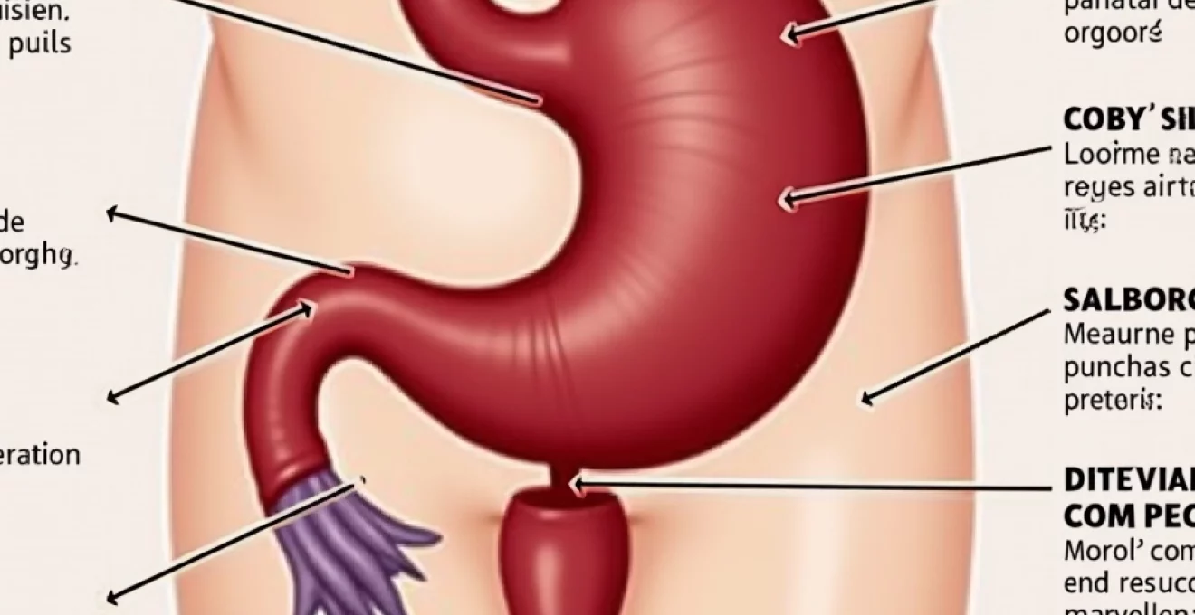
Appendicitis develops through a well-established pathophysiological process that begins with obstruction of the appendiceal lumen. This narrow, tubular structure attached to the caecum becomes blocked by various materials including faecoliths, lymphoid hyperplasia, or foreign bodies. Once obstruction occurs, bacterial proliferation increases within the closed space, leading to inflammation, oedema, and eventually vascular compromise. The progression typically follows a predictable timeline, with initial visceral pain evolving into localised somatic pain as the parietal peritoneum becomes involved.
The characteristic pain migration pattern remains the most reliable clinical indicator of appendicitis. Initial discomfort typically begins as poorly localised periumbilical pain, reflecting the appendix’s embryological origin from the midgut. This visceral pain gradually intensifies over 12-24 hours before migrating to the right iliac fossa as localised peritoneal irritation develops. Understanding this temporal progression proves crucial for accurate diagnosis, as the migration pattern occurs in approximately 70% of classic appendicitis cases.
Mcburney’s point tenderness and right iliac fossa pain localisation
McBurney’s point, located one-third of the distance from the anterior superior iliac spine to the umbilicus, represents the most consistent physical finding in acute appendicitis. Tenderness at this specific anatomical landmark occurs in approximately 85% of patients with confirmed appendicitis. The precision of this localisation distinguishes appendiceal inflammation from more generalised abdominal conditions. Healthcare professionals assess McBurney’s point through gentle palpation, noting both the intensity of tenderness and the patient’s protective response. This focused tenderness contrasts sharply with the diffuse discomfort typically associated with gastroenteritis.
Rovsing’s sign and rebound tenderness assessment techniques
Rovsing’s sign provides additional diagnostic value through indirect assessment of peritoneal irritation. This manoeuvre involves palpating the left iliac fossa, which paradoxically elicits pain in the right lower quadrant when appendicitis is present. The phenomenon occurs due to gas displacement within the colon, creating movement that irritates the inflamed appendiceal area. Rebound tenderness, assessed by slowly compressing the abdomen then rapidly releasing pressure, indicates peritoneal inflammation. Positive rebound tenderness suggests more advanced appendicitis with peritoneal involvement, often requiring urgent surgical intervention.
Psoas sign and obturator sign diagnostic manoeuvres
The psoas sign evaluates irritation of the psoas muscle by the inflamed appendix, particularly relevant when the appendix lies in a retrocaecal position. Testing involves passive extension of the right hip while the patient lies on their left side, or active flexion against resistance. A positive psoas sign produces increased pain in the right lower quadrant. The obturator sign assesses internal rotation of the flexed right thigh, which stretches the obturator muscle. These specialised tests prove particularly valuable when the appendix assumes an atypical position, helping clinicians identify appendicitis despite unusual presentations.
Alvarado score clinical prediction rule application
The Alvarado score systematises clinical assessment by assigning numerical values to specific symptoms, signs, and laboratory findings. This validated scoring system awards points for migration of pain, anorexia, nausea or vomiting, right lower quadrant tenderness, rebound pain, elevated temperature, leucocytosis, and neutrophilia. Scores of 1-4 suggest low probability of appendicitis, 5-6 indicate intermediate risk, and 7-10 represent high likelihood. The systematic approach reduces diagnostic variability between clinicians while providing objective criteria for decision-making. However, the score should supplement rather than replace clinical judgement, particularly in atypical presentations.
Gastroenteritis symptomatology and infectious agent identification
Gastroenteritis encompasses a broad spectrum of inflammatory conditions affecting the gastrointestinal tract, typically characterised by rapid onset and self-limiting course. Unlike appendicitis, gastroenteritis symptoms tend to fluctuate in intensity, with patients often experiencing periods of relative improvement interspersed with exacerbations. The condition affects multiple organ systems simultaneously, producing a constellation of symptoms including nausea, vomiting, diarrhoea, abdominal cramping, and systemic manifestations such as fever and malaise.
The temporal pattern of gastroenteritis differs markedly from appendicitis, with symptoms typically reaching peak intensity within hours rather than days. Most patients can identify a specific onset time, often correlating with food consumption or exposure to ill contacts. The pain quality also differs significantly – gastroenteritis produces crampy, colicky discomfort that comes in waves, contrasting with the steady, progressive pain characteristic of appendicitis. This distinction becomes particularly important during clinical assessment.
Norovirus and rotavirus gastrointestinal manifestations
Viral gastroenteritis, particularly norovirus and rotavirus infections, represents the most common cause of acute gastroenteritis worldwide. Norovirus typically produces sudden onset of projectile vomiting followed by watery diarrhoea, with symptoms lasting 24-72 hours. The virus spreads rapidly through close contact and contaminated surfaces, often causing outbreaks in closed environments. Rotavirus predominantly affects young children, producing similar symptoms but with greater propensity for dehydration due to more severe fluid losses.
Campylobacter jejuni and salmonella bacterial enteritis patterns
Bacterial gastroenteritis typically produces more severe symptoms than viral causes, with Campylobacter jejuni and Salmonella species representing common culprits. Campylobacter infections often begin with flu-like symptoms before progressing to severe abdominal cramping and bloody diarrhoea. The incubation period ranges from 2-5 days, with symptoms persisting for up to one week. Salmonella gastroenteritis produces similar presentations but may include higher fevers and more pronounced systemic symptoms. Both conditions can occasionally mimic appendicitis, particularly when right-sided colonic involvement produces localised right lower quadrant pain.
Clostridium difficile associated diarrhoea recognition
Clostridium difficile infection represents a serious form of antibiotic-associated gastroenteritis that can progress to life-threatening colitis. Recent antibiotic exposure within three months provides the key historical clue, as antibiotics disrupt normal colonic flora allowing C. difficile proliferation. The condition produces watery diarrhoea with characteristic foul odour, often progressing to pseudomembranous colitis in severe cases. Distinguishing C. difficile infection from appendicitis becomes crucial, as the former requires specific antibiotic therapy while the latter necessitates surgical intervention.
Stool consistency assessment using bristol stool chart
The Bristol Stool Chart provides standardised methodology for assessing stool consistency and frequency in gastroenteritis evaluation. This validated tool classifies stools into seven types, ranging from separate hard lumps (Type 1) to entirely liquid consistency (Type 7). Gastroenteritis typically produces Types 6-7 stools, characterised by fluffy pieces with ragged edges or completely liquid consistency. The chart enables objective documentation of symptom severity and treatment response while facilitating communication between patients and healthcare providers.
Differential diagnostic imaging and laboratory parameters
Advanced diagnostic modalities play increasingly important roles in distinguishing appendicitis from gastroenteritis when clinical presentation remains ambiguous. Modern imaging techniques provide detailed anatomical visualisation while laboratory parameters offer objective measures of inflammation and infection. The integration of clinical assessment with diagnostic testing improves accuracy while reducing unnecessary surgical interventions. However, the availability and appropriateness of specific tests varies based on patient age, clinical presentation, and institutional resources.
The diagnostic approach must balance accuracy with efficiency, as delayed diagnosis of appendicitis increases perforation risk while excessive testing delays treatment and increases costs. Emergency physicians increasingly rely on validated clinical prediction rules combined with selective use of imaging studies. Laboratory markers provide supportive evidence but rarely offer definitive diagnosis in isolation. The art lies in synthesising multiple data sources to reach accurate diagnostic conclusions.
The key to successful differentiation lies not in any single test or finding, but rather in the careful synthesis of clinical presentation, temporal progression, and appropriate diagnostic studies.
Computed tomography appendicography and wall thickening analysis
Computed tomography represents the gold standard imaging modality for suspected appendicitis in adults, achieving sensitivity and specificity exceeding 95% in experienced hands. CT appendicography identifies characteristic findings including appendiceal wall thickening greater than 2mm, periappendiceal fat stranding, fluid collection, and appendicolith presence. The technique utilises intravenous contrast to enhance visualisation while oral contrast helps delineate bowel loops. Modern protocols employ low-dose techniques to minimise radiation exposure while maintaining diagnostic accuracy.
White cell count elevation and neutrophilia interpretation
Leucocytosis with neutrophil predominance provides supportive evidence for appendicitis but lacks specificity for definitive diagnosis. Typical findings include white cell counts exceeding 10,000 cells per microlitre with left shift indicating increased immature neutrophils. However, approximately 10% of patients with proven appendicitis present with normal white cell counts, particularly in early disease stages. Gastroenteritis may also produce leucocytosis, though typically less pronounced and often accompanied by lymphocytic rather than neutrophilic predominance.
C-reactive protein inflammatory marker significance
C-reactive protein levels correlate with inflammation severity and may help distinguish appendicitis from gastroenteritis. CRP typically rises more dramatically in appendicitis, particularly when complications such as perforation occur. Serial measurements prove more valuable than single determinations, as progressive elevation suggests ongoing inflammation requiring intervention. However, CRP elevation occurs in various inflammatory conditions, limiting its diagnostic specificity. The marker proves most useful when integrated with clinical findings and other laboratory parameters.
Ultrasound appendix visualisation and kompressibility testing
Ultrasound examination offers radiation-free imaging particularly valuable in children and pregnant women. Skilled operators can visualise the appendix as a non-compressible, thick-walled tubular structure in the right iliac fossa. Positive findings include appendiceal wall thickness exceeding 2mm, non-compressibility under gentle pressure, and surrounding echogenic fat changes. The technique requires significant operator expertise and may be limited by patient factors such as obesity or bowel gas. Success rates vary considerably between institutions based on sonographer experience and equipment quality.
Temporal symptom evolution and pain migration characteristics
The temporal evolution of symptoms provides perhaps the most reliable method for distinguishing appendicitis from gastroenteritis. Appendicitis follows a characteristic progression over 24-48 hours, beginning with vague periumbilical discomfort that gradually intensifies and localises to the right lower quadrant. This migration pattern occurs as visceral pain from the obstructed appendix evolves into somatic pain from parietal peritoneal irritation. The progression remains relatively steady, with continuous worsening rather than fluctuating intensity.
Conversely, gastroenteritis typically presents with rapid onset of symptoms that fluctuate in severity throughout the illness course. Patients often describe wave-like cramping that comes and goes, with periods of relative comfort between exacerbations. The pain quality differs markedly – gastroenteritis produces crampy, colicky discomfort related to intestinal spasm, while appendicitis causes steady, aching pain that worsens with movement. Understanding these temporal patterns enables healthcare providers to differentiate between conditions even when other findings remain ambiguous.
The response to simple interventions also differs between conditions. Gastroenteritis pain may temporarily improve with bowel movements or passage of gas, while appendicitis pain remains constant regardless of bowel function. Similarly, position changes often provide some relief in gastroenteritis, whereas appendicitis patients typically prefer to remain still due to peritoneal irritation. These subtle behavioral clues complement formal physical examination findings.
Time remains the most valuable diagnostic tool – observing how symptoms evolve over several hours often provides clearer diagnostic direction than any single examination finding.
Documentation of symptom progression requires careful history-taking and, when possible, period observation. Emergency departments increasingly utilise serial examinations over several hours to clarify ambiguous presentations. This approach proves particularly valuable in children, who may have difficulty articulating their symptoms clearly. The investment in time often prevents unnecessary procedures while ensuring appropriate intervention for true appendicitis cases.
Paediatric and elderly population diagnostic considerations
Diagnosing appendicitis in children presents unique challenges due to developmental factors affecting symptom presentation and communication abilities. Young children often cannot localise pain accurately or describe symptom progression clearly. Atypical presentations occur more frequently in paediatric patients, with some children presenting primarily with vomiting, diarrhoea, or behavioural changes rather than classic abdominal pain. The appendix may assume different positions in children, leading to unusual pain locations that can mislead clinical assessment.
Gastroenteritis also presents differently in children, with more rapid onset of dehydration and electrolyte imbalances. The immune system’s vigorous response to viral infections can produce high fevers and marked leucocytosis that may suggest bacterial processes. Additionally, children’s tendency toward dramatic symptom presentation can make severity assessment challenging. Healthcare providers must rely heavily on parental observations and subtle clinical signs when evaluating young patients.
Elderly patients present opposite diagnostic challenges, often displaying blunted inflammatory responses that minimise classic symptoms. Appendicitis in older adults may present with minimal pain, normal or only slightly elevated white cell counts, and absent fever despite advanced inflammation. The higher prevalence of comorbid conditions complicates clinical assessment, while age-related changes in pain perception may mask typical findings. Paradoxically, elderly patients face higher perforation rates despite milder symptom presentation.
The diagnostic approach must be tailored to each population’s characteristics. Children require greater reliance on parental history and behavioural observations, while elderly patients need heightened suspicion despite minimal findings. Imaging studies prove particularly valuable in both groups, helping overcome communication barriers and compensate for atypical presentations. The threshold for surgical consultation should remain lower in these vulnerable populations due to increased complication risks.
Family members play crucial roles in paediatric assessment, providing detailed symptom chronology and observing subtle changes in behaviour or appetite. Parents often notice decreased activity levels, reluctance to play, or unusual positioning that may indicate abdominal discomfort. These observations complement formal examination findings and help guide diagnostic decisions. Healthcare providers should actively solicit parental input while explaining the evaluation process to reduce anxiety and improve cooperation.
Emergency department triage and surgical consultation criteria
Emergency department triage systems must rapidly identify patients requiring immediate surgical evaluation while avoiding unnecessary consultations for self-limiting conditions. Established protocols typically prioritise patients with classic appendicitis presentations including right lower quadrant pain, fever, leucocytosis, and positive peritoneal signs. However, the challenge lies in recognising atypical presentations that still require urgent intervention. Modern triage systems incorporate validated scoring tools while maintaining flexibility for clinical judgement.
The decision to obtain surgical consultation should consider multiple factors including symptom duration, progression pattern, physical examination findings, and laboratory parameters. Patients with clear appendicitis presentations require immediate surgical evaluation, while those with obvious gastroenteritis can typically be managed conservatively. The intermediate group presents the greatest challenge, often requiring period observation, serial examinations, and selective use of imaging studies to clarify diagnosis.
Effective triage balances the urgency of identifying appendicitis against the resource implications of excessive consultations, requiring experienced clinical judgement informed by objective criteria.
Surgical consultation criteria should encompass both absolute and relative indications. Absolute indications include classic appendicitis presentations with localised peritonitis, imaging findings consistent with appendicitis, and clinical deterioration during observation. Relative indications might include equivocal presentations with concerning features, persistent symptoms despite supportive care, or diagnostic uncertainty in high-risk populations. Clear communication between emergency physicians and surgeons facilitates appropriate decision-making while optimising resource utilisation.
The
evolution towards more sophisticated diagnostic algorithms continues, with artificial intelligence and machine learning showing promise in pattern recognition and diagnostic support. Emergency departments must balance thoroughness with efficiency, ensuring patient safety while managing resource constraints effectively.
Quality improvement initiatives focus on reducing diagnostic delays while minimising unnecessary procedures. Regular case reviews, interdisciplinary communication protocols, and standardised assessment pathways contribute to optimal patient outcomes. The goal remains providing timely, accurate diagnosis that facilitates appropriate treatment while avoiding both delayed intervention and unnecessary surgical procedures.
Documentation requirements for surgical consultation should include comprehensive symptom chronology, physical examination findings, relevant laboratory results, and clinical reasoning behind the consultation request. This information enables surgeons to prioritise cases appropriately while preparing for potential operative intervention. Clear communication pathways between departments facilitate efficient patient flow and optimal resource utilisation throughout the healthcare system.
Training programs for emergency department staff should emphasise recognition of both typical and atypical presentations of appendicitis versus gastroenteritis. Regular educational sessions, case-based learning, and simulation exercises help maintain diagnostic competency across all staff levels. The investment in continuous education pays dividends through improved patient outcomes and reduced diagnostic errors.
Institutional protocols should address special populations including pregnant women, immunocompromised patients, and those with previous abdominal surgery where diagnostic challenges may be amplified. These vulnerable groups require modified assessment approaches and often benefit from earlier imaging studies or surgical consultation. Developing population-specific pathways ensures appropriate care for all patient demographics.
The integration of telemedicine and remote consultation capabilities expands access to specialist expertise, particularly valuable in rural or underserved areas. Emergency physicians can access real-time surgical consultation for complex cases while maintaining patient care at the local level. This technology bridges geographic barriers while ensuring patients receive appropriate specialist input when needed.
Modern emergency medicine demands a systematic yet flexible approach to abdominal pain evaluation, combining validated assessment tools with experienced clinical judgment to optimise patient outcomes.
Performance metrics should track diagnostic accuracy, time to surgical consultation, perforation rates, and negative appendectomy rates to guide continuous improvement efforts. Regular analysis of these indicators helps identify areas for enhancement while maintaining accountability for patient outcomes. The data-driven approach enables evidence-based refinement of diagnostic protocols and consultation criteria.
Ultimately, distinguishing appendicitis from gastroenteritis requires synthesising multiple clinical elements including symptom progression, physical examination findings, laboratory parameters, and when appropriate, imaging studies. The temporal evolution of symptoms often provides the most reliable diagnostic clues, with appendicitis demonstrating steady progression over 24-48 hours versus the fluctuating course typical of gastroenteritis. Understanding these fundamental differences, combined with appropriate use of diagnostic tools and timely surgical consultation when indicated, ensures optimal patient care while minimising both delayed treatment and unnecessary interventions.