Discovering a ball or lump on the back of your neck can be an unsettling experience that naturally raises concerns about your health. The cervical region, where the neck meets the hairline, is a common location for various types of growths and swellings due to its complex anatomy and frequent exposure to irritants. From simple cysts filled with keratin to benign fatty tumours and reactive lymph nodes responding to infections, the causes of neck lumps are remarkably diverse. Understanding these different conditions can help you recognise when a lump requires immediate medical attention versus when it might be a benign growth that simply needs monitoring. The good news is that the vast majority of neck lumps are non-cancerous and treatable, though proper medical evaluation remains essential for accurate diagnosis and appropriate treatment.
Sebaceous cyst formation and epidermoid cyst development
Cystic growths represent some of the most common causes of lumps on the back of the neck, with sebaceous and epidermoid cysts being particularly prevalent in this anatomical region. These benign formations develop when normal skin structures become blocked or disrupted, leading to the accumulation of various substances beneath the skin’s surface. The neck’s unique environment, characterised by frequent friction from clothing, hair products, and natural oils, creates ideal conditions for cyst formation.
Keratin accumulation in blocked hair follicles
Epidermoid cysts, often mistakenly called sebaceous cysts, form when keratin becomes trapped beneath the skin surface due to blocked hair follicles or damaged skin cells. Keratin , the protein that forms the outer layer of skin and hair, normally sheds naturally but can accumulate when normal skin renewal processes are disrupted. These cysts typically feel firm and moveable under the skin, with a characteristic dome-shaped appearance that may have a small central opening or punctum.
The development of epidermoid cysts on the neck often correlates with poor hygiene practices, excessive sweating, or trauma to hair follicles from shaving or tight clothing. Unlike other types of cysts, epidermoid formations contain a thick, cheese-like substance with a distinctly unpleasant odour when ruptured. While these cysts are generally harmless, they can become infected if bacteria enter through the punctum, leading to pain, redness, and potential complications requiring antibiotic treatment.
Sebaceous gland obstruction and oil retention
True sebaceous cysts, though less common than epidermoid varieties, develop when sebaceous glands become blocked, preventing the normal flow of sebum to the skin surface. These oil-producing glands are particularly abundant around hair follicles on the neck, making this region susceptible to sebaceous cyst formation. The trapped sebum creates a smooth, round lump that feels softer than epidermoid cysts and typically lacks the characteristic punctum.
Hormonal fluctuations, particularly during adolescence or periods of stress, can increase sebum production and contribute to gland obstruction. Sebaceous cyst formation often occurs in individuals with naturally oily skin or those who use heavy hair care products that can clog pores around the hairline. Unlike epidermoid cysts, sebaceous formations rarely become infected but can grow steadily over time if left untreated.
Pilar cyst formation at hair follicle roots
Pilar cysts, also known as trichilemmal cysts, specifically develop around hair follicle roots and are commonly found on the scalp extending down to the nape of the neck. These cysts form when the outer root sheath of hair follicles becomes disrupted, leading to keratin accumulation in a different pattern than epidermoid cysts. Pilar cyst development is strongly linked to genetic factors, with many individuals having a family history of similar growths.
The distinctive characteristic of pilar cysts is their tendency to occur in multiples and their predilection for hair-bearing areas. These formations typically feel firmer than sebaceous cysts and may grow more rapidly than other cystic types. While pilar cysts can become quite large, they rarely become infected due to their location and the type of keratin they contain, which differs from that found in epidermoid cysts.
Genetic predisposition to gardner’s Syndrome-Related cysts
In rare cases, multiple cysts on the neck may be associated with Gardner’s syndrome, a genetic condition that predisposes individuals to developing numerous epidermoid cysts alongside other growths. This autosomal dominant disorder affects approximately 1 in 10,000 people and is characterised by the development of multiple cysts before age 20. Gardner’s syndrome manifestations in the neck region often present as clusters of firm, moveable lumps that may be accompanied by similar growths elsewhere on the body.
The genetic nature of this condition means that family screening may be appropriate when multiple cysts are identified in young individuals. While the cysts themselves are benign, Gardner’s syndrome carries additional health risks that require ongoing medical surveillance, including increased susceptibility to colorectal polyps and other complications that necessitate regular monitoring and preventive care.
Lipoma development and benign adipose tissue growths
Lipomas represent another significant category of benign neck lumps, consisting of mature fat cells that have grown beyond their normal boundaries to form discrete, encapsulated masses. These soft tissue tumours are among the most common benign growths in adults, with the neck being a frequent location due to the abundance of subcutaneous fat in this region.
Lipomas are characterised by their soft, doughy texture and their tendency to move freely under the skin when pressed, distinguishing them from firmer cystic growths or fixed inflammatory masses.
Subcutaneous lipoma formation in cervical region
Subcutaneous lipomas develop just beneath the skin surface and are the most common type found on the back of the neck. These growths typically present as soft, painless lumps that feel somewhat like a small bag of marbles under the skin. The subcutaneous lipoma development process is generally slow, with most growths taking months or years to reach noticeable size.
The exact cause of subcutaneous lipoma formation remains unclear, though research suggests a combination of genetic predisposition and environmental factors may contribute to their development. Age appears to be a significant factor, with most subcutaneous lipomas appearing in individuals between 40 and 60 years old. While these growths are typically cosmetic concerns rather than medical emergencies, they can occasionally interfere with clothing or become tender if they reach substantial size.
Intramuscular lipoma growth within trapezius muscle
Less commonly, lipomas can develop within the muscle tissue itself, particularly within the trapezius muscle that spans the neck and shoulder region. Intramuscular lipomas present differently from their subcutaneous counterparts, often feeling firmer and less mobile due to their integration within muscle fibres. These deeper lipomas may cause discomfort during neck movement or muscle contraction, making them more symptomatic than surface growths.
The diagnosis of intramuscular lipomas often requires imaging studies such as MRI or ultrasound to distinguish them from other soft tissue masses and to determine their relationship to surrounding structures. Intramuscular lipoma management may be more complex than superficial varieties, as surgical removal requires careful attention to preserve muscle function and avoid damage to nearby nerves or blood vessels.
Familial multiple lipomatosis syndrome manifestations
Some individuals develop multiple lipomas throughout their body, including the neck region, as part of familial multiple lipomatosis syndrome. This inherited condition is characterised by the development of numerous lipomas, often beginning in early adulthood and continuing throughout life. The neck is a common site for these multiple growths, which can create significant cosmetic concerns and, in some cases, functional limitations.
The genetic basis of familial multiple lipomatosis involves mutations in genes that regulate fat cell development and growth. Multiple lipomatosis manifestations in the neck region may include dozens of small to medium-sized growths that can merge or cluster together, creating larger, more complex masses that may require specialised surgical approaches for optimal management.
Angiolipoma with vascular component characteristics
Angiolipomas represent a unique variant of lipomatous growths that contain both fat cells and numerous small blood vessels, giving them distinctive characteristics compared to conventional lipomas. These vascular-containing masses are often more tender than typical lipomas and may be sensitive to pressure or temperature changes. The neck region can harbour angiolipomas, particularly in areas where blood supply is abundant.
The vascular component of angiolipomas makes them more prone to pain and tenderness, especially during periods of hormonal fluctuation or temperature extremes. Diagnosis of angiolipomas may require careful clinical examination and sometimes imaging studies to identify the vascular component and distinguish them from other soft tissue masses that might require different treatment approaches.
Lymphadenopathy and reactive lymph node enlargement
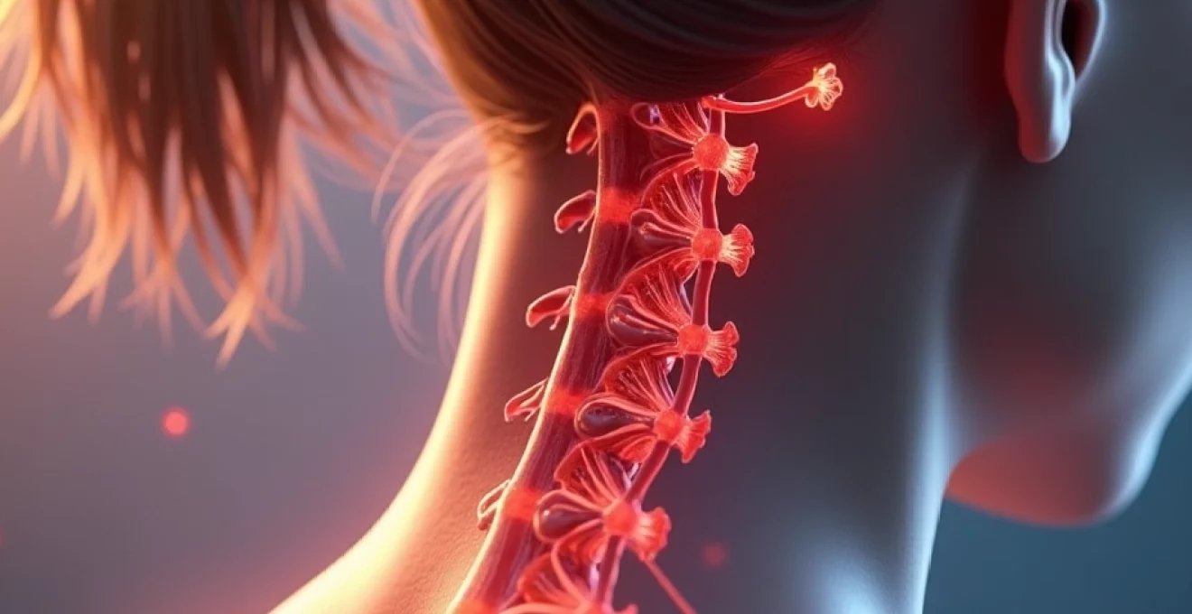
Lymph node enlargement represents one of the most common causes of lumps in the neck region, with the cervical lymph node chains being particularly responsive to infections and inflammatory processes affecting the head and neck area. The human neck contains numerous lymph node groups, including the superficial cervical chain along the external jugular vein, the deep cervical chain following the internal jugular vein, and the posterior cervical nodes along the posterior border of the sternocleidomastoid muscle. When these nodes become enlarged, they create palpable lumps that can vary significantly in size, consistency, and associated symptoms.
Reactive lymphadenopathy occurs when lymph nodes respond to antigenic stimulation, whether from infectious agents, inflammatory conditions, or other immune system triggers. The enlargement process involves increased cellular activity within the node, including proliferation of lymphocytes, macrophages, and other immune cells working to process and respond to foreign substances. This cellular activity causes the node to swell beyond its normal size, creating the characteristic lumps that individuals notice during self-examination or routine medical assessment.
The characteristics of enlarged lymph nodes can provide important diagnostic clues about their underlying cause.
Benign reactive lymph nodes typically feel soft to rubbery, are mobile under the skin, and may be tender to palpation, particularly during acute infectious processes.
In contrast, lymph nodes affected by malignant processes often feel hard, fixed to surrounding tissues, and are generally painless, though these distinctions are not absolute and require professional medical evaluation for accurate assessment.
Common infectious causes of cervical lymphadenopathy include viral upper respiratory infections, bacterial pharyngitis, dental infections, and mononucleosis. Each of these conditions can cause characteristic patterns of node enlargement, with viral infections often causing bilateral, tender enlargement, while bacterial infections may produce more asymmetric, painful swelling. The temporal relationship between symptom onset and node enlargement often provides additional diagnostic information, with acute infections typically causing rapid node enlargement that resolves as the infection clears.
More serious causes of lymph node enlargement include lymphomas, metastatic cancer from head and neck primary tumours, and systemic conditions such as sarcoidosis or autoimmune disorders. Malignant lymphadenopathy often presents with nodes that are progressively enlarging, painless, and may be associated with constitutional symptoms such as unexplained weight loss, night sweats, or persistent fatigue. The evaluation of persistent lymph node enlargement requires careful clinical assessment and may necessitate additional testing including imaging studies or tissue biopsy for definitive diagnosis.
Inflammatory conditions and infectious processes
Inflammatory and infectious processes affecting the posterior neck region can create various types of lumps and swellings that may be mistaken for other conditions. These processes range from superficial skin infections to deeper soft tissue inflammations, each presenting with characteristic features that can help distinguish them from non-inflammatory causes of neck lumps. The area where the hairline meets the neck is particularly susceptible to these conditions due to its exposure to hair care products, friction from clothing, and the presence of numerous hair follicles that can serve as entry points for bacterial pathogens.
Folliculitis represents one of the most common inflammatory conditions affecting the neck area, occurring when hair follicles become infected with bacteria, typically Staphylococcus aureus. This condition creates small, red, tender bumps around hair follicles that may progress to form larger, more significant lumps if the infection spreads to surrounding tissues. Follicular inflammation in the neck region is often exacerbated by practices such as frequent shaving, use of occlusive hair products, or wearing tight clothing that creates friction against the skin.
Carbuncles and furuncles (boils) represent more severe forms of follicular infection that can create substantial lumps on the back of the neck. These deep skin infections involve multiple hair follicles and surrounding tissue, creating painful, red, swollen masses that may eventually develop central cores of purulent material. The development of carbuncles is often associated with predisposing factors such as diabetes, immunosuppression, or poor hygiene practices that allow bacterial colonisation to progress to deeper tissue invasion.
Hidradenitis suppurativa, though more commonly affecting areas such as the axillae and groin, can occasionally involve the neck region, particularly in areas where apocrine glands are present. This chronic inflammatory condition creates recurrent, painful nodules that may progress to form interconnected sinus tracts and scarring. The inflammatory nature of hidradenitis suppurativa can create complex masses that may be mistaken for other types of growths, requiring careful clinical evaluation and sometimes specialised testing for accurate diagnosis.
Infectious processes affecting deeper structures in the neck, such as lymphangitis or cellulitis, can create diffuse swelling and induration that may be perceived as lumps by affected individuals. These conditions often present with characteristic signs of inflammation including redness, warmth, tenderness, and sometimes systemic symptoms such as fever and malaise. The rapid onset and inflammatory characteristics of these conditions help distinguish them from slower-developing, non-inflammatory causes of neck lumps.
Muscular and soft tissue abnormalities
Muscular and soft tissue abnormalities in the neck region can create palpable lumps that may be mistaken for other types of growths, particularly when they develop gradually or result from chronic conditions. The complex muscular anatomy of the neck, including the trapezius, levator scapulae, splenius muscles, and deeper cervical musculature, provides numerous opportunities for the development of muscular knots, trigger points, and other abnormalities that can present as discrete lumps or areas of induration.
Myofascial trigger points represent one of the most common muscular causes of perceived lumps in the neck region. These hyperirritable spots within skeletal muscle fibres can develop following muscle overuse, poor posture, or acute injury, creating discrete areas of muscle tension that feel like firm nodules under the skin. Trigger point development in the upper trapezius and cervical paraspinal muscles is particularly common in individuals who spend extended periods working at computers or engaging in activities that require sustained neck positioning.
The characteristics of myofascial trigger points help distinguish them from other causes of neck lumps: they are typically tender to palpation, may reproduce familiar pain patterns when pressed, and often demonstrate a characteristic “twitch response” when stimulated. Unlike cysts or lipomas, trigger points are not truly separate structures but rather areas of altered muscle physiology that can be modified through appropriate treatment approaches including massage, stretching, and trigger point release techniques.
Muscle hernias, though relatively uncommon in the neck region, can occasionally occur where muscle fibres protrude through defects in the overlying fascia, creating palpable lumps that may become more prominent during muscle contraction. These herniation can result from congenital fascial defects or acquired weakness following trauma or surgical procedures. Muscular herniation in the neck typically presents as a soft, reducible mass that becomes more prominent during activities that increase muscle tension.
Fibromatosis and other proliferative soft tissue conditions can affect the neck region, creating firm, sometimes irregular masses within the muscular or fascial layers. These conditions involve abnormal proliferation of fibrous tissue and can present challenges in diagnosis due to their similarity to other soft tissue masses. The evaluation of suspected fibromatous conditions often requires imaging studies and sometimes tissue sampling to distinguish them from other soft tissue abnormalities and to guide appropriate treatment decisions.
Vascular abnormalities, including arteriovenous malformations and venous ectasias, can occasionally present as palpable masses in the neck region. These vascular anomalies may demonstrate characteristic features such as pulsation, change in size with position, or audible bruits that help distinguish them from other causes of neck lumps. The evaluation of suspected vascular abnormalities typically requires specialised imaging studies to define the anatomy and flow characteristics of the lesion, information that is essential
for proper treatment planning and to avoid potential complications during any required interventions.
Contractures and adhesive conditions affecting the cervical fascia can create areas of thickening and restriction that may be perceived as lumps or masses, particularly following previous surgery, radiation therapy, or significant trauma to the neck region. These fibrotic changes typically develop gradually and may be associated with functional limitations including reduced range of motion or discomfort during neck movement. Fascial contracture development often requires multidisciplinary management approaches that may include physical therapy, manual treatments, and sometimes surgical release procedures to restore normal tissue mobility and function.
The evaluation of muscular and soft tissue abnormalities in the neck requires careful clinical assessment to distinguish between various conditions that can present with similar physical findings. Healthcare providers typically assess factors such as the relationship of lumps to muscle contraction, changes in size or consistency with positioning, and the presence of associated functional limitations. Advanced imaging studies, including MRI or ultrasound, may be necessary to fully characterise soft tissue abnormalities and guide appropriate treatment decisions, particularly when conservative management approaches have not provided adequate symptom relief or when the diagnosis remains uncertain after initial clinical evaluation.