Nasal breathing difficulties affect millions of people worldwide, yet the underlying causes often remain misunderstood or misdiagnosed. Two of the most prevalent conditions causing chronic nasal obstruction are deviated septum and nasal polyps, both of which can significantly impact quality of life through persistent congestion, reduced sense of smell, and sleep disturbances. While these conditions may present with similar symptoms, they differ fundamentally in their anatomical origins, pathophysiology, and treatment approaches. Understanding these distinctions is crucial for patients seeking effective relief and for healthcare professionals providing accurate diagnoses. The complexity of sinonasal anatomy means that both conditions can coexist, making comprehensive evaluation essential for optimal treatment outcomes.
Anatomical structure and pathophysiology of deviated septum
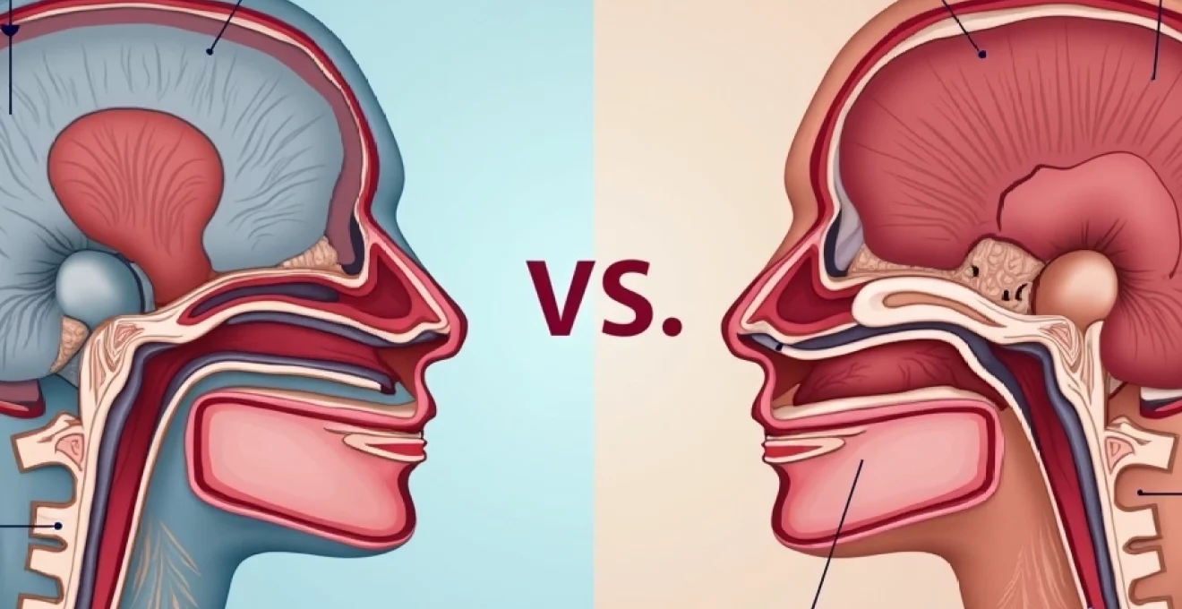
Nasal septum cartilage and bone composition
The nasal septum represents a sophisticated anatomical structure comprising both cartilaginous and bony components that divide the nasal cavity into distinct chambers. The anterior portion consists primarily of quadrangular cartilage, which provides flexibility and structural support during nasal development and growth. This cartilaginous component extends from the nasal tip to approximately the middle third of the nasal cavity, where it transitions to the bony septum.
The posterior bony septum includes the perpendicular plate of the ethmoid bone superiorly and the vomer bone inferiorly. These structures articulate with the nasal spine of the maxilla and palatine bones, creating a complex three-dimensional framework. The intricate junction points between these anatomical components often become areas of weakness where septal deviations commonly develop, particularly during periods of rapid growth or following trauma.
Types of septal deviations: C-Shape, S-Shape, and localised deformities
Septal deviations manifest in various morphological patterns, each presenting unique challenges for nasal airflow and surgical correction. C-shaped deviations represent the most common pattern, where the septum curves uniformly to one side, creating asymmetric nasal chambers with one significantly narrowed passage. This configuration typically results in consistent unilateral obstruction that patients often describe as being able to breathe through only one nostril.
S-shaped deviations present greater complexity, featuring multiple curves that create alternating areas of narrowing on both sides of the nasal cavity. These deviations often develop following significant trauma or during periods of asymmetric growth, resulting in bilateral symptoms that may vary depending on nasal cycle patterns and environmental factors such as allergen exposure or viral infections.
Localised septal deformities, including septal spurs and isolated ridges, represent focal areas of bony or cartilaginous overgrowth. These discrete abnormalities can cause significant symptoms despite their limited size, particularly when they contact the lateral nasal wall or middle turbinate, creating areas of mucosal irritation and potential secondary inflammation.
Impact on nasal airflow dynamics and turbulent flow patterns
Septal deviations fundamentally alter the laminar flow patterns essential for optimal nasal function, creating areas of turbulent airflow that reduce the efficiency of nasal breathing, humidification, and filtration. The nasal cavity’s natural architecture promotes smooth, laminar airflow that allows for proper contact between inspired air and the nasal mucosa, facilitating warming, humidification, and particulate filtration.
When septal deviations narrow specific areas of the nasal passage, air velocity increases significantly in these regions, similar to how water flows faster through a narrow section of a river. This increased velocity creates turbulent flow patterns that generate the sensation of nasal obstruction and can lead to mucosal drying and irritation. The disrupted airflow also reduces the effectiveness of the nasal cycle, a normal physiological process where nasal passages alternately congest and decongest throughout the day.
Research indicates that even minor septal deviations can reduce nasal airflow efficiency by up to 40%, significantly impacting respiratory comfort and sleep quality.
Congenital versus acquired septal deviation mechanisms
Congenital septal deviations result from abnormal intrauterine development or birth trauma, affecting approximately 20% of newborns to some degree. During foetal development, the nasal septum forms through the fusion of multiple embryonic structures, and any disruption in this complex process can result in developmental abnormalities. Birth trauma, particularly during difficult deliveries requiring forceps or vacuum assistance, can also cause immediate septal displacement.
Acquired septal deviations typically result from nasal trauma occurring after birth, with childhood injuries being particularly significant due to the ongoing growth and development of nasal structures. Sports injuries, falls, and motor vehicle accidents represent common causes, with the degree of deviation often correlating with the severity and direction of the initial trauma. Interestingly , many acquired deviations worsen over time as the nose continues to grow and age, making early detection and monitoring important for optimal outcomes.
Nasal polyps formation and histopathological characteristics
Inflammatory polyp development in chronic rhinosinusitis
Nasal polyps develop as a consequence of chronic mucosal inflammation, representing the end result of prolonged inflammatory cascade activation within the sinonasal mucosa. The pathogenesis involves complex interactions between environmental triggers, genetic predisposition, and immune system dysfunction that ultimately leads to excessive tissue proliferation and oedema formation. Unlike the structural abnormalities seen in septal deviation, polyp formation represents an active inflammatory process.
The inflammatory environment characteristic of chronic rhinosinusitis creates ideal conditions for polyp development through sustained elevation of inflammatory mediators including interleukins, leukotrienes, and prostaglandins. These mediators promote increased vascular permeability, tissue oedema, and fibroblast proliferation, resulting in the characteristic grape-like growths that can progressively enlarge to obstruct nasal passages completely.
Environmental factors such as allergen exposure, bacterial biofilms, and fungal elements contribute to the perpetuation of the inflammatory cycle. The damaged epithelial barrier allows increased penetration of irritants and allergens, creating a self-perpetuating cycle of inflammation and tissue remodelling that characterises chronic rhinosinusitis with nasal polyposis.
Eosinophilic and neutrophilic polyp phenotypes
Modern understanding of nasal polyps recognises distinct inflammatory phenotypes that influence both clinical presentation and treatment response. Eosinophilic polyps represent the most common Western phenotype, characterised by predominant eosinophil infiltration and associated with type 2 immune responses. These polyps typically present with severe anosmia, extensive sinonasal involvement, and higher recurrence rates following surgical treatment.
The eosinophilic phenotype often associates with comorbid asthma and aspirin sensitivity, creating a challenging clinical syndrome requiring multimodal management approaches. Elevated tissue eosinophilia correlates with increased disease severity and reduced response to conventional corticosteroid therapy, necessitating consideration of newer targeted biological therapies.
Neutrophilic polyps, more prevalent in certain geographic regions and populations, demonstrate predominant neutrophil infiltration and often associate with bacterial colonisation or infection. This phenotype typically shows better response to antimicrobial therapy and may present with more purulent rhinorrhoea compared to the eosinophilic variant.
Antrochoanal polyps versus ethmoidal polyposis distribution
The anatomical distribution of nasal polyps provides important diagnostic and therapeutic insights, with distinct patterns correlating with different underlying pathophysiology. Antrochoanal polyps represent solitary inflammatory masses originating from the maxillary sinus, extending through the middle meatus into the nasal cavity and potentially reaching the nasopharynx. These polyps typically affect younger patients and demonstrate unilateral presentation with characteristic imaging findings.
Ethmoidal polyposis, conversely, represents diffuse polyp formation throughout the ethmoid air cell system, often extending into multiple sinus cavities simultaneously. This pattern typically associates with chronic rhinosinusitis and presents bilaterally, though asymmetric involvement is common. The extensive nature of ethmoidal polyposis often requires more aggressive surgical management and demonstrates higher recurrence rates compared to isolated antrochoanal polyps.
Aspirin-exacerbated respiratory disease (AERD) association
Aspirin-Exacerbated Respiratory Disease represents a distinct clinical syndrome affecting approximately 7% of adults with asthma and up to 15% of patients with nasal polyposis. This condition, also known as Samter’s triad, encompasses the classic combination of nasal polyps, asthma, and aspirin sensitivity, though the complete triad may not always be present at initial diagnosis.
The pathophysiology of AERD involves dysregulation of arachidonic acid metabolism, particularly affecting the cyclooxygenase and lipoxygenase pathways. Aspirin and other NSAIDs inhibit cyclooxygenase activity, leading to increased leukotriene production and subsequent intense inflammatory responses. Patients with AERD typically develop extensive nasal polyposis with characteristic eosinophilic infiltration and demonstrate aggressive disease progression requiring intensive medical and surgical management.
AERD patients exhibit recurrence rates of up to 80% following surgical treatment, highlighting the importance of comprehensive medical management and patient education regarding aspirin avoidance.
Clinical presentation and differential symptomatology
Unilateral versus bilateral nasal obstruction patterns
The pattern of nasal obstruction provides crucial diagnostic information for distinguishing between septal deviation and nasal polyposis. Septal deviations typically produce predictable unilateral obstruction corresponding to the narrowed nasal passage, though patients may experience alternating symptoms due to the natural nasal cycle. The obstructed side often remains consistent, with patients able to identify which nostril provides better airflow.
Nasal polyps, particularly in their early stages, may also present with unilateral symptoms, especially in cases of antrochoanal polyps or asymmetric ethmoidal disease. However, as polyp disease progresses, bilateral involvement becomes increasingly common, often resulting in complete nasal obstruction that patients describe as being unable to breathe through either nostril.
The temporal pattern of obstruction also differs significantly between these conditions. Septal deviation typically produces stable, consistent symptoms that may worsen during upper respiratory infections or seasonal allergies but generally remain predictable. Polyp-related obstruction often demonstrates progressive worsening over months to years, with periods of partial improvement during anti-inflammatory treatment followed by symptom recurrence.
Anosmia and hyposmia: distinguishing olfactory dysfunction
Olfactory dysfunction represents one of the most significant quality-of-life impacts associated with sinonasal disease, yet the mechanisms differ substantially between septal deviation and nasal polyposis. Septal deviation primarily causes conductive olfactory loss through mechanical obstruction of airflow to the olfactory cleft, the narrow space high in the nasal cavity where olfactory receptors are concentrated.
In contrast, nasal polyps cause both conductive and sensorineural olfactory dysfunction. The conductive component results from physical obstruction of the olfactory cleft by polyp tissue, while the sensorineural component develops from chronic inflammation affecting the olfactory neuroepithelium directly. This dual mechanism explains why patients with nasal polyps often experience more severe and persistent olfactory dysfunction compared to those with septal deviation alone.
The reversibility of olfactory dysfunction also differs between these conditions. Patients with septal deviation may notice immediate improvement in smell function following successful surgical correction, as the mechanical obstruction is relieved. However, polyp patients often experience gradual, incomplete recovery of olfactory function even after successful treatment, reflecting the irreversible damage to olfactory neurons caused by chronic inflammation.
Rhinorrhoea characteristics and Post-Nasal drip variations
The characteristics of nasal discharge provide valuable diagnostic information for differentiating between septal deviation and polyp-related pathology. Patients with isolated septal deviation typically report minimal rhinorrhoea, with any discharge generally being clear and related to concurrent conditions such as allergic rhinitis or viral upper respiratory infections.
Nasal polyps, conversely, often produce characteristic thick, tenacious mucus that patients describe as difficult to clear. The inflammatory process underlying polyp formation results in altered mucus composition with increased viscosity and reduced ciliary clearance. This leads to the typical complaints of persistent post-nasal drip, frequent throat clearing, and the sensation of mucus accumulation in the posterior nasal cavity.
The colour and consistency of discharge can also provide diagnostic clues. Clear, watery discharge suggests allergic or non-allergic rhinitis, while thick, discoloured secretions may indicate secondary bacterial infection, particularly common in patients with significant polyp disease where impaired drainage predisposes to bacterial colonisation and biofilm formation.
Sleep-disordered breathing and snoring manifestations
Both septal deviation and nasal polyps significantly impact sleep quality through mechanisms affecting upper airway patency and breathing patterns. The relationship between nasal obstruction and sleep-disordered breathing reflects the complex interdependence of nasal breathing, oral breathing compensation, and upper airway stability during sleep.
Septal deviation contributes to snoring and sleep apnoea through increased nasal resistance, which promotes mouth breathing and reduces the natural splinting effect of nasal airflow on the upper airway. The resulting oral breathing during sleep leads to posterior tongue positioning and increased collapsibility of the pharyngeal tissues, potentially contributing to obstructive sleep apnoea development or exacerbation.
Nasal polyps often produce more severe sleep disruption due to the typically bilateral and progressive nature of obstruction. Complete nasal obstruction forces obligate mouth breathing, which not only increases snoring but also leads to morning dry mouth, throat irritation, and daytime fatigue. The inflammatory mediators associated with polyp disease may also directly affect sleep architecture and quality independent of the mechanical obstruction.
Advanced diagnostic imaging and endoscopic assessment
High-resolution CT scan interpretation using Lund-Mackay scoring
High-resolution computed tomography represents the gold standard for evaluating sinonasal anatomy and pathology, providing detailed visualisation of both septal abnormalities and polyp distribution. Modern CT protocols utilise thin-section imaging with multiplanar reconstructions, allowing precise assessment of septal deviation patterns, contact points, and the extent of sinus involvement in polyp disease.
The Lund-Mackay scoring system provides standardised quantification of chronic rhinosinusitis severity based on CT findings. This system assigns scores from 0-2 for each sinus group (maxillary, anterior ethmoids, posterior ethmoids, sphenoid, and frontal) and the ostiomeatal complex, with total scores ranging from 0-24. Higher scores correlate with more extensive disease and often indicate need for surgical intervention.
CT imaging also reveals important anatomical variants that may predispose to chronic rhinosinusitis or complicate surgical treatment. Concha bullosa, paradoxic middle turbinate curvature, and Haller cells represent common variants that can contribute to ostiomeatal complex obstruction and subsequent inflammatory disease development.
Nasal endoscopy findings: septal spurs and polyp grading
Nasal endoscopy provides direct visualisation of sinonasal anatomy and pathology, offering superior assessment compared to anterior rhinoscopy alone. Modern flexible and rigid endoscopic techniques allow detailed examination of the nasal cavity, middle meatus, and sphenoethmoidal recess, areas crucial for understanding disease patterns and planning treatment approaches.
Septal deviations are readily apparent on endoscopic examination, with particular attention paid to contact points between deviated septal segments and the lateral nasal wall. These contact points often develop secondary mucosal changes including ulceration, crusting, or inflammatory polyp formation, highlighting the complex relationship between anatomical abnormalities and inflammatory disease.
Polyp grading systems utilise endoscopic findings to quantify disease severity and track treatment response. The most commonly used system grades polyps from 0-4 based on size and extent, with Grade 0 representing no polyps, Grade 1 indicating small polyps not reaching the inferior turbinate, and Grade 4 representing massive polyposis completely obstructing the nasal cavity.
Acoustic rhinometry and rhinomanometry measurements
Objective measurement
of nasal airflow provides objective assessment of respiratory function that complements subjective symptom reporting and endoscopic findings. Acoustic rhinometry utilises sound reflection technology to measure nasal cross-sectional areas and volumes, providing detailed information about the geometric properties of the nasal cavity that influence airflow resistance.
This non-invasive technique identifies the location and severity of nasal narrowing with remarkable precision, distinguishing between septal deviation and mucosal swelling as causes of obstruction. Acoustic rhinometry demonstrates particular value in monitoring treatment response and surgical outcomes, as measurements can be repeated easily without patient discomfort or radiation exposure.
Rhinomanometry measures nasal airflow resistance by recording pressure and flow relationships during controlled breathing manoeuvres. Active anterior rhinomanometry represents the most commonly used technique, measuring airflow through one nostril while the opposite nostril is occluded. This approach provides quantitative data about unilateral nasal function that correlates well with patient-reported symptoms and quality of life measures.
Combined acoustic rhinometry and rhinomanometry assessments demonstrate up to 85% correlation with patient-reported symptom severity, making these tools valuable for treatment planning and outcome assessment.
MRI applications in complex sinonasal pathology
Magnetic resonance imaging provides superior soft tissue contrast compared to CT scanning, making it particularly valuable for evaluating complex sinonasal pathology where differentiation between inflammatory tissue, retained secretions, and potential neoplasms is crucial. T2-weighted imaging demonstrates excellent visualisation of mucosal thickening and fluid collections, while T1-weighted sequences with gadolinium enhancement help identify areas of active inflammation or vascular malformations.
The ability of MRI to distinguish between different types of soft tissue makes it invaluable when evaluating patients with extensive polyposis where malignancy must be excluded. Fungal sinusitis, particularly invasive forms, demonstrates characteristic signal patterns on MRI that help guide urgent treatment decisions. Additionally, MRI provides excellent visualisation of intracranial complications, making it essential when evaluating patients with suspected complications of chronic sinusitis.
Advanced MRI techniques including diffusion-weighted imaging and dynamic contrast enhancement offer emerging applications in sinonasal disease evaluation. These methods may help differentiate between inflammatory and neoplastic processes with greater accuracy than conventional imaging, potentially reducing the need for tissue sampling in selected cases.
Surgical management: septoplasty versus functional endoscopic sinus surgery
The surgical management of septal deviation and nasal polyposis requires distinctly different approaches, reflecting the fundamental differences in underlying pathophysiology and anatomical involvement. Septoplasty focuses on correcting structural abnormalities to restore normal nasal anatomy and airflow patterns, while functional endoscopic sinus surgery (FESS) addresses the inflammatory disease process and restores normal sinus drainage and ventilation.
Septoplasty techniques have evolved significantly with the introduction of endoscopic visualisation and powered instrumentation. Modern approaches emphasise preservation of nasal support structures while achieving maximal functional improvement. The procedure typically involves careful elevation of mucoperichondrial and mucoperiosteal flaps, selective removal or repositioning of deviated cartilage and bone, and meticulous reconstruction to maintain nasal tip support and external nasal appearance.
Functional endoscopic sinus surgery for nasal polyposis requires a more extensive approach, addressing both the polyp disease and the underlying chronic rhinosinusitis. Complete polypectomy combined with wide opening of affected sinuses allows for improved ventilation, drainage, and post-operative medical management. The extent of surgery depends on disease severity, with procedures ranging from limited polypectomy and middle meatal antrostomy to complete sphenoethmoidectomy with frontal sinus procedures.
Combined procedures addressing both septal deviation and nasal polyposis are frequently necessary, as anatomical abnormalities often contribute to the development and perpetuation of inflammatory disease. Sequential or simultaneous surgical approaches must be carefully planned to optimise functional outcomes while minimising complications. The decision-making process requires thorough evaluation of disease severity, patient symptoms, and individual anatomical considerations.
Post-operative care protocols differ significantly between these procedures, reflecting the distinct healing processes involved. Septoplasty patients typically require nasal packing or splints for structural support during initial healing, while FESS patients benefit from intensive nasal irrigation and topical corticosteroid therapy to prevent adhesion formation and recurrent inflammation. Understanding these differences is crucial for achieving optimal surgical outcomes and patient satisfaction.
Long-term prognosis and recurrence prevention strategies
The long-term prognosis for patients with septal deviation and nasal polyposis differs substantially, reflecting the distinct nature of these conditions and their response to treatment interventions. Septal deviation, being primarily a structural abnormality, typically demonstrates excellent long-term outcomes following successful surgical correction, with patient satisfaction rates exceeding 80% at five-year follow-up when appropriate surgical techniques are employed.
Recurrence of septal deviation following septoplasty is relatively uncommon, occurring in fewer than 10% of patients and usually resulting from inadequate initial correction, post-operative trauma, or continued nasal growth in younger patients. The key to preventing septal deviation recurrence lies in thorough initial surgical correction, preservation of nasal support structures, and patient education regarding activity restrictions during the healing period.
Nasal polyposis presents greater challenges regarding long-term management and recurrence prevention, with recurrence rates ranging from 15-40% depending on underlying disease phenotype and patient compliance with medical therapy. Eosinophilic polyposis demonstrates particularly high recurrence rates, often requiring revision surgery within five years of initial treatment. The chronic inflammatory nature of the disease necessitates lifelong medical management even after successful surgical intervention.
Comprehensive recurrence prevention strategies for nasal polyposis include aggressive topical corticosteroid therapy, allergen avoidance when relevant, treatment of concurrent asthma, and consideration of systemic anti-inflammatory medications in severe cases. Newer biological therapies targeting specific inflammatory pathways show promise for reducing recurrence rates in patients with severe, recalcitrant disease.
Recent studies indicate that patients receiving combination therapy including high-volume saline irrigation, topical corticosteroids, and appropriate biological agents demonstrate recurrence rates below 20% at three-year follow-up.
Patient education plays a crucial role in long-term success, particularly for those with nasal polyposis who must understand the chronic nature of their condition and the importance of ongoing medical management. Regular follow-up appointments allow for early detection of disease recurrence and timely intervention before symptoms become severe. The development of objective monitoring tools including patient-reported outcome measures and endoscopic scoring systems helps optimise long-term care strategies.
Environmental factors significantly influence long-term outcomes in both conditions. Patients with septal deviation may experience symptom fluctuation related to seasonal allergies or viral infections, but these typically resolve without affecting the structural correction achieved through surgery. However, patients with nasal polyposis require ongoing attention to environmental triggers, occupational exposures, and concurrent medical conditions that may influence disease activity and treatment response.
The emergence of precision medicine approaches offers exciting prospects for improving long-term outcomes in nasal polyposis management. Genetic testing for specific inflammatory markers, assessment of individual medication metabolism patterns, and personalised biological therapy selection may revolutionise treatment approaches and significantly improve recurrence prevention strategies in the coming years.