Small round rashes appearing on the neck present a diagnostic challenge that requires careful evaluation of morphological characteristics, distribution patterns, and underlying pathophysiology. The cervical region’s unique anatomical features, including increased moisture retention, frequent contact with clothing and accessories, and exposure to various environmental factors, make it particularly susceptible to diverse dermatological conditions. Understanding the spectrum of potential aetiologies—from infectious agents to allergic reactions—enables healthcare providers and patients alike to identify appropriate treatment pathways and prevent potential complications.
The neck’s proximity to hair follicles, sebaceous glands, and lymphatic drainage systems creates a complex microenvironment where multiple pathological processes can manifest as seemingly similar round rash presentations. Early recognition of distinguishing features becomes crucial for implementing targeted therapeutic interventions and avoiding the progression of treatable conditions into more severe manifestations.
Dermatological classification of small round neck rashes
Professional dermatological assessment relies heavily on systematic classification of cutaneous lesions based on their morphological characteristics, distribution patterns, and temporal evolution. Small round rashes on the neck encompass a broad spectrum of primary and secondary skin lesions that require methodical evaluation to establish accurate diagnoses.
Primary morphological characteristics and visual identification
Primary lesions represent the initial manifestation of pathological processes without secondary changes from scratching, infection, or treatment. Papules appear as solid, raised lesions less than 1 centimetre in diameter, often presenting as the earliest sign of inflammatory or infectious conditions affecting the cervical region. These elevated lesions may demonstrate varying degrees of induration, surface texture, and pigmentation depending on the underlying pathophysiology.
Macules represent flat, circumscribed areas of colour change without elevation or depression, frequently observed in viral exanthems or early stages of contact dermatitis. The distinction between papular and macular presentations significantly influences diagnostic considerations and treatment selection. Vesicles, characterised by fluid-filled cavities less than 1 centimetre in diameter, often indicate viral infections or acute allergic reactions requiring immediate therapeutic intervention.
Papular versus vesicular lesion differentiation
The differentiation between papular and vesicular lesions requires careful inspection under adequate lighting conditions, as this distinction fundamentally alters diagnostic possibilities and treatment approaches. Papular lesions typically maintain their solid consistency throughout the disease course, while vesicular lesions may progress through various stages of fluid accumulation, rupture, and crust formation.
Vesicular eruptions often present with surrounding erythema and may demonstrate umbilication or central depression, particularly in viral conditions such as molluscum contagiosum. The temporal evolution of these lesions provides valuable diagnostic information, as vesicles typically develop rapidly and may resolve spontaneously or progress to pustular or crusted stages.
Erythematous and Non-Erythematous presentation patterns
Erythematous presentations indicate active inflammation with increased vascular permeability and blood flow to affected tissues. The degree and pattern of erythema often correlate with the severity and nature of the underlying pathological process. Non-erythematous lesions may suggest chronic conditions, post-inflammatory changes, or certain infectious processes that do not elicit significant inflammatory responses.
The assessment of erythema requires consideration of individual skin phototypes, as inflammatory changes may appear less pronounced or present with different colour characteristics in darker skin tones. Purple, brown, or grey discolouration may represent inflammatory processes in individuals with higher melanin content, necessitating adapted diagnostic criteria.
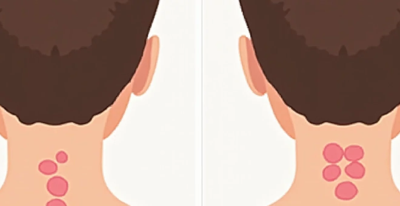
Size parameters and clustered distribution analysis
Precise measurement of individual lesions and assessment of their spatial distribution provide crucial diagnostic information. Small round rashes typically measure between 2-10 millimetres in diameter, though size variations may occur based on the duration and severity of the condition. Clustered distributions often suggest infectious aetiologies or contact sensitisation patterns.
Linear arrangements may indicate contact with specific irritants or allergens, while scattered random distributions might suggest systemic causes or arthropod bite reactions. The bilateral symmetry or asymmetric presentation further narrows diagnostic possibilities and guides appropriate investigative approaches.
Infectious aetiologies behind cervical round rash formation
Infectious causes represent a significant proportion of small round rashes affecting the cervical region, encompassing bacterial, viral, and fungal pathogens that exploit the neck’s unique anatomical characteristics. The warm, moist environment created by clothing, hair, and natural skin folds provides optimal conditions for microbial proliferation and subsequent inflammatory responses.
Folliculitis and staphylococcus aureus bacterial colonisation
Bacterial folliculitis primarily results from Staphylococcus aureus invasion of hair follicles, creating characteristic perifollicular pustules that may appear as small round lesions on the neck. The condition typically develops following minor trauma from shaving, tight clothing, or excessive friction, allowing bacterial penetration through compromised follicular barriers.
Early-stage folliculitis presents as small erythematous papules centred around hair follicles, progressing to pustular lesions within 24-48 hours. The neck’s high concentration of sebaceous glands and hair follicles makes it particularly susceptible to recurrent episodes, especially in individuals with underlying conditions that compromise immune function or skin barrier integrity.
Herpes simplex virus reactivation and vesicular eruptions
Herpes simplex virus reactivation can manifest on the neck as clusters of small vesicular lesions, though this presentation remains less common than facial or genital involvement. The characteristic progression from tingling sensations to vesicle formation, followed by ulceration and crusting, typically occurs over 7-10 days.
Prodromal symptoms including burning, itching, or hypersensitivity often precede visible lesion development by 12-24 hours. Secondary bacterial infection may complicate the healing process, particularly when vesicles rupture and expose underlying tissue to environmental contaminants. Recurrent episodes typically demonstrate shorter duration and reduced severity compared to primary infections.
Tinea corporis fungal infection manifestations
Dermatophyte infections affecting the neck typically present as expanding annular lesions with raised, scaly borders and central clearing. However, early-stage infections may appear as small round patches without the characteristic ring formation, complicating initial diagnosis and treatment decisions.
The neck’s exposure to moisture from perspiration and occlusion from clothing creates favourable conditions for fungal proliferation. Tinea corporis lesions often demonstrate asymmetric distribution and may coalesce to form larger irregular patches. The presence of satellite lesions around the primary site suggests active fungal spread requiring extended treatment duration.
Molluscum contagiosum viral papule development
Molluscum contagiosum produces characteristic dome-shaped papules with central umbilication, though early lesions may appear as simple round bumps without obvious central depression. The viral infection primarily affects immunocompromised individuals and children, with neck involvement occurring through direct contact or autoinoculation.
Individual lesions typically measure 2-5 millimetres in diameter and may persist for months to years without treatment. The waxy, pearl-like appearance and firm consistency help differentiate molluscum from other papular conditions. Spontaneous resolution eventually occurs in immunocompetent individuals, though treatment may accelerate clearance and reduce transmission risk.
Contact dermatitis and allergic reaction mechanisms
Contact dermatitis represents one of the most frequent causes of round rash formation on the neck, resulting from direct exposure to irritants or allergens through jewellery, cosmetics, clothing, and occupational materials. The pathophysiology involves either immediate irritant reactions or delayed-type hypersensitivity responses mediated by T-lymphocyte activation.
Understanding the distinction between irritant and allergic contact dermatitis becomes crucial for implementing appropriate avoidance strategies and selecting optimal therapeutic interventions.
Nickel sensitivity from jewellery and metal accessories
Nickel represents the most common contact allergen worldwide, with neck involvement frequently resulting from prolonged contact with jewellery, clothing fasteners, and metal accessories. The allergic response typically develops within 12-72 hours of exposure, manifesting as erythematous, vesicular, or papular lesions in areas of direct metal contact.
Chronic nickel dermatitis may present as thickened, lichenified skin with persistent scaling and hyperpigmentation. The geometric pattern of lesions often mirrors the shape and size of the offending metal object, providing valuable diagnostic clues. Sweat and friction increase nickel release from metal alloys, explaining the higher prevalence during warm weather or physical activity.
Fragrance allergen reactions from cosmetic products
Fragrance components in perfumes, colognes, lotions, and personal care products frequently cause allergic contact dermatitis on the neck due to direct application or transfer from hands. The complex mixture of natural and synthetic aromatic compounds makes identification of specific allergens challenging without formal patch testing procedures.
Essential oils, including those marketed as “natural” alternatives, contain potent sensitising compounds such as limonene , linalool, and eugenol. The neck’s increased sensitivity to fragrance allergens results from its thin skin, high vascular density, and frequent occlusion by clothing and hair. Cross-reactivity between related fragrance chemicals may complicate avoidance strategies.
Textile dye and fabric softener induced responses
Textile-related contact dermatitis on the neck commonly results from formaldehyde-releasing preservatives, disperse dyes, and fabric treatment chemicals. New clothing items pose particular risks due to higher concentrations of processing chemicals that gradually diminish with repeated washing.
Fabric softeners and laundry additives containing quaternary ammonium compounds may cause delayed allergic reactions, particularly in areas where clothing maintains close skin contact. Para-phenylenediamine and related dye components in dark-coloured clothing represent significant allergens that may cause persistent dermatitis despite proper laundering procedures.
Occupational contact dermatitis from chemical exposure
Workplace exposure to industrial chemicals, cleaning agents, and processing materials may cause contact dermatitis extending to the neck region through direct contact or transfer from contaminated hands and clothing. Healthcare workers, food service employees, and industrial workers demonstrate increased susceptibility due to frequent chemical exposure.
Latex proteins, disinfectants, and preservatives represent common occupational allergens affecting neck skin through airborne particles or indirect transfer mechanisms. Occupational dermatitis often requires comprehensive exposure assessment and workplace modification to achieve sustainable improvement. The chronic nature of occupational exposure may lead to persistent sensitisation requiring long-term management strategies.
Seborrhoeic dermatitis and inflammatory skin conditions
Seborrhoeic dermatitis affecting the neck typically presents as well-demarcated, erythematous patches with adherent yellowish scales, though early manifestations may appear as small round inflammatory lesions before developing characteristic scaling patterns. The condition results from complex interactions between Malassezia yeast overgrowth, altered sebum composition, and individual inflammatory responses.
The neck’s sebaceous gland density and frequent occlusion by clothing and hair create optimal conditions for Malassezia proliferation and subsequent inflammatory cascade activation. Stress, hormonal fluctuations, and immunosuppression may exacerbate symptoms and trigger acute flares. The chronic relapsing nature of seborrhoeic dermatitis requires long-term management strategies incorporating both therapeutic interventions and lifestyle modifications.
Differential diagnosis from other inflammatory conditions requires careful evaluation of scale characteristics, distribution patterns, and response to antifungal treatments. The presence of similar lesions in other seborrhoeic areas, including the scalp, face, and chest, supports the diagnostic impression and guides treatment selection.
Secondary bacterial infection may complicate seborrhoeic dermatitis, particularly when excessive scratching compromises skin barrier function. The development of pustular lesions or rapid symptom progression warrants antimicrobial therapy in addition to standard anti-inflammatory treatments. Chronic inflammation may lead to post-inflammatory hyperpigmentation or hypopigmentation, requiring additional therapeutic considerations.
Heat rash and sweat gland dysfunction disorders
Heat rash, or miliaria, represents a spectrum of conditions resulting from sweat gland occlusion and subsequent inflammatory responses. The neck’s predisposition to heat retention, moisture accumulation, and clothing friction makes it particularly susceptible to various forms of miliaria during warm weather or periods of increased perspiration.
Miliaria crystallina presents as small, clear vesicles without surrounding inflammation, representing the most superficial form of sweat gland obstruction. These fragile lesions typically rupture easily and resolve rapidly with cooling and reduced perspiration. The absence of inflammatory changes distinguishes this condition from infectious or allergic causes of vesicular eruptions.
Miliaria rubra, commonly known as “prickly heat,” manifests as small erythematous papules and vesicles accompanied by intense itching and burning sensations. The inflammatory response results from sweat leakage into deeper skin layers following ductal obstruction. Secondary infection may occur when excessive scratching introduces pathogenic bacteria through compromised skin barriers.
The prevention of heat rash relies primarily on environmental modifications to reduce perspiration and improve air circulation around affected areas, making lifestyle counselling an essential component of management strategies.
Miliaria profunda represents the deepest form of sweat gland obstruction, presenting as flesh-coloured papules without associated vesiculation. This condition primarily affects individuals with chronic heat exposure and may progress to anhidrotic heat exhaustion due to reduced sweating capacity. The neck’s involvement in extensive miliaria profunda may indicate significant thermal regulation compromise requiring immediate medical attention.
Chronic heat exposure and repeated episodes of miliaria may lead to permanent sweat gland dysfunction and altered thermoregulatory capacity. The development of compensatory hyperhidrosis in unaffected body regions represents an adaptive response that may create new sites of heat rash formation, necessitating comprehensive management approaches.
Diagnostic assessment and clinical evaluation protocols
Comprehensive diagnostic assessment of small round neck rashes requires systematic evaluation incorporating detailed history-taking, physical examination, and selective laboratory investigations. The initial clinical encounter should focus on establishing the temporal relationship between symptom onset and potential triggering factors, including new medications, cosmetic products, jewellery, or environmental exposures.
Morphological assessment begins with careful inspection under adequate lighting conditions, documenting lesion characteristics, distribution patterns, and any associated secondary changes such as excoriation, lichenification, or pigmentary alterations. Digital photography may facilitate monitoring of treatment response and provide valuable documentation for specialist referrals when indicated.
The patient’s occupational history, recreational activities, and personal care routines provide essential contextual information for identifying potential causative factors. Recent travel, new pet exposure, or changes in living environment may suggest infectious aetiologies requiring specific antimicrobial treatments. Family history of atopic conditions or autoimmune diseases may influence diagnostic considerations and treatment selection.
Laboratory investigations should be selectively employed based on clinical suspicion rather than routine screening, as most small round neck rashes can be diagnosed through careful clinical assessment and therapeutic trials.
Potassium hydroxide preparation and fungal culture remain valuable diagnostic tools when dermatophyte infection is suspected, particularly in cases with characteristic scaling or failure to respond to standard treatments. Bacterial culture and sensitivity testing may guide antibiotic selection in cases of suspected secondary bacterial infection or atypical presentations.
Patch testing represents the gold standard for identifying specific contact allergens in cases of suspected allergic contact dermatitis. The procedure requires temporary discontinuation of topical corticosteroids and careful patient preparation to ensure reliable results. Standard patch test series should be supplemented with relevant occupational or personal allergens based on individual exposure patterns.
Dermoscopy, when available, may provide additional morphological details that aid in distinguishing between similar-appearing conditions. The identification of specific dermoscopic features such as follicular involvement, scale patterns, or vascular arrangements can significantly enhance diagnostic accuracy and reduce the need for more invasive procedures.
Biopsy should be reserved for cases with atypical presentations, failure to respond to appropriate treatment, or suspicion of malignant transformation. The
selection of an appropriate biopsy site and technique requires consideration of lesion characteristics and suspected pathology to maximise diagnostic yield while minimising cosmetic impact.
The integration of clinical findings with patient history and selective diagnostic testing enables accurate diagnosis in the vast majority of cases presenting with small round neck rashes. Therapeutic trials using topical treatments may serve both diagnostic and therapeutic purposes, particularly when distinguishing between inflammatory and infectious conditions with overlapping clinical presentations.
Patient education regarding proper skin care, allergen avoidance, and when to seek follow-up care forms an essential component of the diagnostic encounter. Clear documentation of findings, suspected diagnoses, and treatment plans facilitates continuity of care and enables appropriate specialist referrals when conservative management fails to achieve satisfactory outcomes.
Follow-up scheduling should account for the expected timeline of improvement based on the suspected diagnosis and treatment approach. Most inflammatory conditions show initial improvement within 1-2 weeks of appropriate treatment, while infectious causes may require longer treatment durations to prevent recurrence and ensure complete pathogen eradication.
The systematic approach to diagnosing small round neck rashes combines clinical expertise with selective use of diagnostic tools to achieve accurate diagnosis while avoiding unnecessary procedures and reducing healthcare costs.
Emergency referral criteria include rapidly spreading rashes with systemic symptoms, signs of severe infection such as cellulitis or lymphangitis, or suspected drug reactions with mucosal involvement. These situations require immediate medical attention to prevent serious complications and ensure appropriate management of potentially life-threatening conditions.
Dermatology referral should be considered for recurrent episodes despite appropriate treatment, atypical presentations that don’t respond to standard therapies, or when patch testing is required to identify specific contact allergens. Chronic conditions requiring long-term management may benefit from specialist input to optimise treatment strategies and prevent complications.
The role of telemedicine in evaluating skin conditions continues to expand, though limitations exist in assessing texture, depth, and subtle colour variations that may influence diagnostic accuracy. High-quality digital photography with appropriate lighting and multiple angles can enhance remote consultation effectiveness when in-person evaluation is not immediately available.