Hernias represent one of the most fascinating yet concerning aspects of human anatomy, demonstrating how seemingly robust bodily structures can develop unexpected weaknesses. While most people associate hernias exclusively with the groin or abdomen, the reality is far more complex and extensive. These anatomical disruptions can occur at numerous locations throughout the human body, each presenting unique challenges for diagnosis, treatment, and long-term management. Understanding where hernias can develop provides crucial insight into preventive measures and helps individuals recognise potentially serious symptoms before complications arise. The human body contains multiple fascial planes, muscle layers, and natural openings that can become sites of herniation under the right circumstances, making hernia awareness essential for maintaining optimal health.
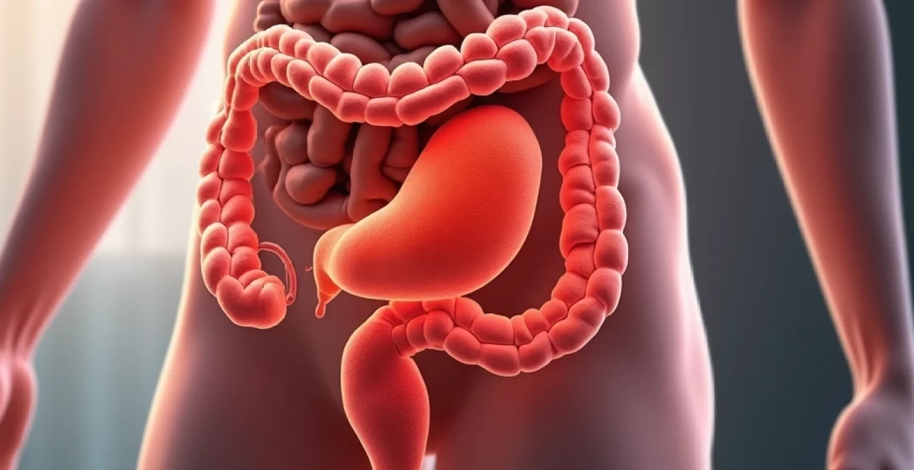
Anatomical distribution of hernia types throughout the human body
The human body contains numerous potential sites for hernia development, far beyond the commonly recognised abdominal wall locations. These anatomical vulnerabilities exist wherever fascial planes meet, natural openings occur, or tissue weakness develops over time. Hernia formation follows predictable patterns based on anatomical structure, with certain locations experiencing higher incidence rates due to inherent structural characteristics or functional demands placed upon specific body regions.
Research indicates that approximately 75% of all hernias occur in the inguinal region, making this the most prevalent location for herniation. However, the remaining 25% of cases span across multiple anatomical sites, including the diaphragm, umbilical region, surgical incision sites, and various rare locations throughout the torso. Each anatomical region presents distinct risk factors, symptoms, and treatment approaches, requiring specialised understanding for optimal patient outcomes.
The distribution of hernia types varies significantly based on demographic factors, with age, gender, and occupational hazards influencing both location and likelihood of development. Men experience inguinal hernias at rates eight to ten times higher than women, whilst women show increased susceptibility to femoral and umbilical herniation patterns. This gender-based variation reflects fundamental anatomical differences in pelvic structure, muscle development, and hormonal influences on connective tissue strength.
Abdominal wall hernias: inguinal, femoral, and umbilical classifications
Abdominal wall hernias represent the most commonly encountered hernia types, accounting for the vast majority of surgical repairs performed annually. Inguinal hernias occur when abdominal contents protrude through the inguinal canal, a natural passage in the lower abdominal wall that allows the spermatic cord in men and the round ligament in women to pass from the abdomen to the external genitalia. These hernias can be classified as direct or indirect, depending on their relationship to the inferior epigastric vessels and their pathway through the abdominal wall.
Femoral hernias develop when tissue pushes through the femoral canal, located just below the inguinal ligament. These hernias are particularly concerning due to their high risk of strangulation, as the narrow femoral ring provides limited space for herniated contents to expand. Women experience femoral hernias more frequently than men, particularly following pregnancy or significant weight changes that alter pelvic anatomy and increase intra-abdominal pressure.
Umbilical hernias occur at the site of the former umbilical cord attachment, where incomplete closure of the umbilical ring creates a potential weakness. In adults, these hernias often develop following pregnancy, significant weight gain, or chronic increases in abdominal pressure. The umbilical region’s central location and rich blood supply generally provide favourable conditions for surgical repair, though large defects may require complex reconstruction techniques.
Diaphragmatic hernias: hiatal and congenital diaphragmatic defects
The diaphragm, a crucial respiratory muscle separating the thoracic and abdominal cavities, contains several natural openings that can become sites of herniation. Hiatal hernias represent the most common diaphragmatic hernia type, occurring when portions of the stomach migrate through the oesophageal hiatus into the chest cavity. These hernias are classified as sliding (Type I) or paraesophageal (Types II-IV), with each classification presenting distinct anatomical configurations and clinical implications.
Sliding hiatal hernias involve upward migration of the gastroesophageal junction, whilst paraesophageal hernias feature herniation of the gastric fundus alongside a normally positioned gastroesophageal junction. Large paraesophageal hernias can accommodate significant portions of the stomach, potentially leading to gastric volvulus, a life-threatening condition requiring emergency surgical intervention.
Congenital diaphragmatic hernias occur during foetal development when the diaphragm fails to form properly, leaving openings through which abdominal organs can enter the chest cavity. These defects most commonly occur on the left side through the foramen of Bochdalek, though right-sided and anterior defects can also develop. The timing and extent of herniation significantly impact foetal lung development, often requiring immediate postnatal surgical repair and intensive respiratory support.
Incisional hernias following surgical interventions
Surgical incisions create intentional disruptions in the abdominal wall that must heal properly to maintain structural integrity. When this healing process fails or becomes compromised, incisional hernias can develop at the surgical site. These hernias occur in approximately 10-15% of abdominal operations, with higher rates observed following emergency procedures, wound infections, or operations performed in patients with compromised healing capacity.
The development of incisional hernias reflects complex interactions between surgical technique, patient factors, and postoperative care. Factors increasing incisional hernia risk include obesity, diabetes, smoking, immunosuppressive medications, and postoperative wound complications. The size and location of the original incision also influence hernia development, with larger incisions and those crossing muscle fibres at unfavourable angles showing increased susceptibility.
Modern surgical techniques emphasise proper fascial closure methods and selective use of prophylactic mesh reinforcement in high-risk patients. These approaches have demonstrated significant reductions in incisional hernia rates, particularly when combined with optimised perioperative care protocols addressing modifiable risk factors such as glycemic control and smoking cessation.
Rare herniation sites: obturator, lumbar, and spigelian locations
Beyond the commonly recognised hernia locations, numerous rare anatomical sites can develop herniation under specific circumstances. Obturator hernias occur through the obturator foramen in the pelvis, typically affecting elderly women with chronic illness and significant weight loss. These hernias often present with vague groin pain and may cause compression of the obturator nerve, leading to characteristic medial thigh pain known as the Howship-Romberg sign.
Lumbar hernias develop through defects in the posterior abdominal wall, either at the superior lumbar triangle (Grynfeltt-Lesshaft triangle) or the inferior lumbar triangle (Petit triangle). These hernias can be congenital or acquired, with acquired forms often following trauma, infection, or surgical procedures in the lumbar region. The deep location and complex anatomy of lumbar hernias frequently make diagnosis challenging, often requiring advanced imaging for confirmation.
Spigelian hernias occur along the semilunar line, lateral to the rectus abdominis muscle, where the aponeuroses of the internal oblique and transversus abdominis muscles meet. These hernias typically remain small and may be difficult to detect on physical examination, earning them the designation “hidden hernias.” Despite their inconspicuous nature, Spigelian hernias carry significant risk for incarceration due to their narrow neck and rigid fascial margins.
Pathophysiology of fascial weakness and tissue protrusion mechanisms
Understanding the fundamental mechanisms underlying hernia development requires examination of the complex interplay between tissue strength, mechanical forces, and biological factors that influence fascial integrity. The pathophysiology of herniation involves progressive weakening of connective tissue structures that normally contain intra-abdominal contents within their appropriate anatomical compartments. This process typically develops over time, though acute herniation can occur following sudden increases in intra-abdominal pressure or traumatic tissue disruption.
The development of hernia involves a cascade of cellular and molecular events that compromise the structural integrity of fascial planes. These changes begin at the microscopic level with alterations in collagen synthesis and degradation patterns, progressing to macroscopic tissue failure when mechanical stresses exceed the reduced tensile strength of weakened tissues. The process is influenced by genetic predisposition, age-related changes, environmental factors, and various disease states that affect connective tissue metabolism.
Research has identified several key pathways involved in hernia development, including altered matrix metalloproteinase activity, changes in collagen type ratios, and disrupted fibroblast function. These molecular changes create a progressive cycle of tissue weakening that predisposes affected areas to herniation when exposed to increased mechanical stress. Understanding these mechanisms has led to improved surgical techniques and the development of biocompatible materials designed to support long-term tissue integrity.
Collagen degradation and connective tissue failure patterns
Collagen represents the primary structural protein providing tensile strength to fascial tissues, with alterations in collagen metabolism playing a central role in hernia development. Normal fascial tissue contains predominantly Type I collagen, which provides superior tensile strength, whilst herniated tissues often show increased ratios of Type III collagen, a less robust structural protein. This shift in collagen composition significantly reduces tissue strength and predisposes to progressive expansion under mechanical stress.
Matrix metalloproteinases (MMPs) regulate collagen turnover through controlled degradation of existing collagen fibres, allowing for tissue remodelling and repair. In hernia development, dysregulated MMP activity leads to excessive collagen breakdown without corresponding increases in synthesis, creating a net loss of structural integrity. Specific MMP subtypes, particularly MMP-2 and MMP-9, show elevated activity in herniated tissues, contributing to the progressive nature of fascial defects.
The spatial organisation of collagen fibres also influences tissue strength, with normal fascial planes exhibiting organised, interwoven fibre patterns that distribute mechanical forces efficiently. Herniated tissues demonstrate disorganised collagen architecture with reduced cross-linking between fibres, compromising the tissue’s ability to withstand multidirectional stresses. This architectural disruption often extends beyond the visible hernia defect, explaining the increased recurrence rates observed when surgical repairs fail to address the broader area of tissue compromise.
Intra-abdominal pressure dynamics and herniation risk factors
Intra-abdominal pressure represents a critical factor in hernia development, with both acute pressure spikes and chronic pressure elevation contributing to fascial failure. Normal intra-abdominal pressure ranges from 5-15 mmHg at rest, but can increase dramatically during activities such as coughing, straining, or heavy lifting. These pressure increases create outward forces on the abdominal wall that must be contained by fascial structures to maintain anatomical integrity.
Chronic conditions that persistently elevate intra-abdominal pressure significantly increase hernia risk through repeated mechanical stress on fascial tissues. Obesity, chronic obstructive pulmonary disease, benign prostatic hyperplasia, and chronic constipation all contribute to sustained pressure elevation that accelerates fascial weakening over time. The cumulative effect of these repeated stress cycles eventually exceeds the tissue’s capacity for repair and regeneration.
The relationship between pressure and herniation follows predictable biomechanical principles, with tissue failure occurring when applied forces exceed the material strength of fascial structures. This relationship explains why hernia development often accelerates following specific triggering events, such as severe coughing episodes or acute increases in physical activity. Understanding these pressure dynamics has informed surgical techniques that aim to distribute forces more effectively and reduce recurrence rates through improved tissue reinforcement strategies.
Congenital versus acquired fascial defects
Hernia development can result from either congenital anatomical variations or acquired tissue weakening, with each category presenting distinct characteristics and treatment considerations. Congenital fascial defects result from incomplete embryological development of abdominal wall structures, creating inherent weak points that may not manifest clinically until later in life when mechanical stresses exceed the compromised tissue’s capacity.
The most common congenital defect leading to adult herniation involves incomplete closure of the processus vaginalis during foetal development. This embryological structure normally obliterates by birth, but persistent patency creates a potential pathway for inguinal herniation. Similarly, incomplete umbilical ring closure can predispose to umbilical hernia development, particularly when combined with factors that increase intra-abdominal pressure.
Acquired fascial defects develop through progressive tissue weakening caused by various factors including aging, mechanical stress, inflammation, and metabolic disturbances. These defects often occur at sites of natural anatomical weakness, such as the inguinal canal or areas where vessels and nerves penetrate fascial planes. The distinction between congenital and acquired defects has important implications for surgical planning, as congenital defects may require different repair strategies to address the underlying anatomical variation.
Age-related changes in tissue elasticity and strength
Aging produces profound changes in connective tissue composition and mechanical properties that significantly influence hernia susceptibility. Age-related collagen changes include progressive loss of elastin fibres, increased cross-linking that reduces tissue flexibility, and decreased synthesis of new collagen to replace damaged fibres. These changes collectively reduce the tissue’s ability to accommodate mechanical stress without failure.
The aging process affects not only the quantity but also the quality of collagen production, with older tissues producing collagen with altered structural properties. Advanced glycation end products accumulate in aged tissues, creating excessive cross-links that make collagen fibres brittle and prone to fracture under stress. This age-related brittleness explains the increased hernia incidence observed in elderly populations, even in the absence of obvious predisposing factors.
Cellular senescence in fibroblasts further compounds age-related tissue weakness by reducing the capacity for tissue repair and regeneration. Senescent fibroblasts produce less collagen and secrete inflammatory mediators that can accelerate tissue degradation. This cellular aging process creates a progressive cycle of tissue weakening that continues throughout life, emphasising the importance of early recognition and appropriate treatment of fascial defects before they progress to clinically significant herniation.
Clinical manifestations across different anatomical regions
The clinical presentation of hernias varies dramatically depending on their anatomical location, size, and relationship to surrounding structures. Understanding these location-specific manifestations is crucial for accurate diagnosis and appropriate treatment planning. Symptoms can range from completely asymptomatic presentations discovered incidentally during routine examinations to life-threatening emergencies requiring immediate surgical intervention.
Groin hernias typically present with visible bulging that becomes more prominent with increased intra-abdominal pressure from coughing, straining, or standing. Patients often describe a dragging sensation or mild discomfort that worsens throughout the day and improves with rest or when lying down. The ability to reduce the hernia by gently pushing the contents back into the abdomen is a reassuring sign indicating that the hernia is not incarcerated or strangulated.
Hiatal hernias present with entirely different symptom profiles, primarily affecting digestive function rather than causing visible external changes. Gastroesophageal reflux symptoms including heartburn, regurgitation, and difficulty swallowing are common presentations, whilst larger hernias may cause chest pain that can mimic cardiac conditions. The symptoms often worsen after meals or when lying flat, reflecting the mechanical displacement of stomach contents into the chest cavity.
Hernia symptoms should never be ignored, as they can progress rapidly from mild discomfort to life-threatening complications requiring emergency surgical intervention.
Incisional hernias frequently present as bulging at previous surgical sites, particularly noticeable when patients perform activities that increase abdominal pressure. The bulge may be tender to touch and can vary in size depending on the time of day and activity level. Pain is often more prominent than with other hernia types, possibly due to adhesions between herniated contents and surrounding scar tissue from previous operations.
Emergency presentations occur when hernias become incarcerated or strangulated, leading to acute severe pain, nausea, vomiting, and inability to reduce the hernia. These symptoms require immediate medical evaluation, as delayed treatment can result in bowel ischemia, perforation, and life-threatening sepsis. Recognition of these warning signs is essential for preventing serious complications and optimising patient outcomes.
Diagnostic imaging modalities for Multi-Site hernia detection
Modern diagnostic imaging has revolutionised hernia detection and characterisation, particularly for cases where clinical examination alone may be insufficient. The choice of imaging modality depends on the suspected hernia location, patient factors, and the clinical question being addressed. Each imaging technique offers unique advantages and limitations that must be considered when developing diagnostic strategies for complex
or atypical hernia presentations.
Ultrasound imaging serves as the primary diagnostic tool for many hernia evaluations, offering real-time visualisation of tissue movement and hernia contents. This non-invasive modality excels at detecting superficial hernias, particularly inguinal and femoral varieties, while allowing dynamic assessment during Valsalva manoeuvres. High-frequency transducers can identify even small fascial defects and distinguish between different types of groin hernias based on their anatomical relationships to surrounding structures.
Computed tomography (CT) scanning provides superior detail for complex abdominal wall hernias and cases where multiple hernia sites are suspected. CT imaging can detect occult hernias not apparent on clinical examination, characterise hernia contents, and identify complications such as bowel obstruction or strangulation. The cross-sectional nature of CT allows comprehensive evaluation of the entire abdominal wall, making it particularly valuable for planning surgical repairs of large or complex defects.
Magnetic resonance imaging (MRI) offers exceptional soft tissue contrast without radiation exposure, making it ideal for evaluating sports hernias and other subtle fascial injuries. MRI can detect early fascial tears before they progress to frank herniation, allowing for preventive interventions. The multiplanar capabilities of MRI provide detailed anatomical information essential for planning complex reconstructive procedures, particularly in cases involving previous failed repairs or extensive scar tissue.
Advanced imaging techniques have reduced diagnostic uncertainty in hernia evaluation from over 20% to less than 5%, significantly improving surgical planning and patient outcomes.
Specialised imaging protocols have been developed for specific hernia types, such as dynamic MRI for pelvic floor hernias and contrast-enhanced studies for hiatal hernia assessment. These targeted approaches maximise diagnostic yield while minimising unnecessary radiation exposure and examination time. The integration of artificial intelligence in imaging interpretation shows promise for further improving diagnostic accuracy and identifying subtle findings that might escape human observation.
Surgical repair techniques for location-specific hernias
The surgical approach to hernia repair has evolved dramatically over recent decades, with technique selection now based on hernia location, size, patient factors, and surgeon expertise. Modern hernia surgery emphasises tension-free repairs using synthetic or biological mesh materials to reinforce weakened fascial planes. The goal extends beyond simple defect closure to restoration of normal anatomical function and prevention of recurrence through biomechanically sound reconstruction techniques.
Location-specific considerations play a crucial role in surgical planning, as each anatomical region presents unique challenges and opportunities. Groin hernias benefit from well-established repair techniques with excellent long-term outcomes, whilst complex abdominal wall defects may require innovative approaches combining multiple surgical principles. The surgeon must consider not only the immediate hernia but also the potential for future herniation at adjacent sites when planning the repair strategy.
Patient factors significantly influence technique selection, with age, comorbidities, activity level, and previous surgical history all affecting the optimal approach. Minimally invasive techniques offer advantages in appropriate candidates, including reduced postoperative pain, faster recovery, and lower infection rates. However, open repairs remain important tools in the surgical armamentarium, particularly for large defects or cases with significant scar tissue from previous operations.
Tension-free mesh repair methods: lichtenstein and TEP approaches
The Lichtenstein tension-free mesh repair revolutionised inguinal hernia surgery by eliminating the high recurrence rates associated with traditional tissue-based repairs. This technique involves placement of a synthetic mesh patch over the posterior inguinal canal, secured with interrupted sutures to surrounding tissues. The mesh provides immediate reinforcement and serves as a scaffold for tissue ingrowth, creating a durable repair that can withstand normal physiological stresses.
Technical aspects of the Lichtenstein repair require careful attention to anatomical landmarks and proper mesh positioning to avoid nerve entrapment and ensure adequate coverage of potential weakness areas. The lateral border of the mesh must extend beyond the anterior superior iliac spine, whilst the medial border should overlap the pubic tubercle by at least 2 centimetres. Proper mesh tailoring and fixation technique are critical for optimal outcomes, with specific suturing patterns designed to accommodate the spermatic cord whilst maintaining secure fixation.
The totally extraperitoneal (TEP) repair approaches inguinal hernias through the preperitoneal space, avoiding entry into the peritoneal cavity. This technique offers the advantage of simultaneous bilateral repair when indicated and may reduce postoperative pain compared to open methods. TEP repair requires advanced laparoscopic skills and thorough understanding of preperitoneal anatomy, as critical structures including major blood vessels and nerves lie in close proximity to the operative field.
Long-term outcomes data demonstrate excellent results for both techniques, with recurrence rates below 2% in experienced hands. Patient selection criteria help determine the optimal approach, with TEP repair particularly advantageous for bilateral hernias, recurrent hernias after open repair, and patients requiring rapid return to physical activity. The learning curve for TEP repair is longer than for Lichtenstein technique, emphasising the importance of proper training and experience in achieving optimal outcomes.
Laparoscopic techniques: TAPP and robotic-assisted procedures
The transabdominal preperitoneal (TAPP) repair provides excellent visualisation of the entire myopectineal orifice, allowing identification and repair of multiple hernia types during a single procedure. This technique involves entering the peritoneal cavity laparoscopically, creating a peritoneal flap, and placing mesh over the entire area of potential weakness. TAPP repair offers superior visualisation compared to TEP techniques but requires peritoneal closure to prevent bowel adhesions to the mesh.
Robotic-assisted hernia repair represents the latest evolution in minimally invasive techniques, offering enhanced visualisation through high-definition 3D optics and improved dexterity through articulating instruments. The robotic platform facilitates precise dissection and suturing in confined spaces, potentially improving outcomes for complex repairs. However, increased operative time and costs must be weighed against potential benefits when selecting patients for robotic repair.
Patient selection for laparoscopic techniques considers factors including hernia size, bilateral presentation, previous surgical history, and patient comorbidities. Contraindications include inability to tolerate general anaesthesia, extensive intra-abdominal adhesions, and certain medical conditions affecting pneumoperitoneum tolerance. The surgeon’s experience level also influences patient selection, as these techniques require significant training to achieve proficiency and optimal outcomes.
Comparative studies demonstrate equivalent or superior outcomes for laparoscopic techniques compared to open repairs, with reduced postoperative pain, faster recovery, and lower wound infection rates. However, rare but serious complications such as bowel injury and major vessel injury can occur, emphasising the importance of proper patient selection and surgeon training. Long-term recurrence rates appear comparable between laparoscopic and open techniques when performed by experienced surgeons.
Open repair strategies for complex anatomical locations
Complex anatomical locations often require open surgical approaches due to limited access for laparoscopic techniques or the need for extensive tissue mobilisation and reconstruction. Large incisional hernias, lumbar hernias, and certain hiatal hernias benefit from open techniques that allow direct visualisation and manipulation of tissues. Component separation techniques enable closure of large defects by mobilising native tissues whilst reducing tension on repair sutures.
The Rives-Stoppa repair technique addresses large ventral hernias through a retromuscular approach, placing mesh in the preperitoneal space behind the rectus muscles. This technique provides excellent mesh integration whilst maintaining the rectus muscles’ function and blood supply. The retromuscular position protects the mesh from intra-abdominal contents whilst utilising the strong posterior rectus sheath for secure fixation.
Biological mesh materials play important roles in contaminated fields or cases where synthetic mesh may be contraindicated. These materials, derived from human, porcine, or bovine sources, provide temporary scaffolding whilst allowing native tissue ingrowth and remodelling. While biological meshes have higher resorption rates compared to synthetic alternatives, they offer advantages in infected fields or cases requiring contact with bowel contents.
Complex repairs often require multidisciplinary approaches involving plastic surgeons, particularly when extensive tissue loss necessitates flap reconstruction. Free tissue transfers or regional flaps can provide vascularised tissue coverage for large defects whilst allowing simultaneous hernia repair. These advanced techniques require careful preoperative planning and experienced surgical teams but can achieve excellent functional and aesthetic outcomes in challenging cases.
Complications and recurrence patterns by anatomical site
Hernia repair complications vary significantly based on anatomical location, repair technique, and patient factors, requiring location-specific understanding for optimal prevention and management. Immediate postoperative complications include bleeding, infection, and organ injury, whilst long-term issues encompass chronic pain, mesh-related problems, and hernia recurrence. Complication rates have decreased substantially with improved surgical techniques and materials, but remain important considerations in surgical planning and patient counselling.
Groin hernia repairs demonstrate excellent overall safety profiles, with major complication rates below 1% in most series. Chronic pain represents the most significant long-term concern, affecting 5-10% of patients following inguinal hernia repair. This pain typically results from nerve entrapment or mesh-related inflammatory responses and can significantly impact quality of life. Careful attention to anatomical landmarks and judicious mesh fixation techniques help minimise these complications.
Ventral and incisional hernia repairs carry higher complication rates due to larger operative fields and more complex anatomy. Surgical site infections occur in 5-15% of cases, with higher rates observed in patients with obesity, diabetes, or previous infections. The use of prophylactic antibiotics, optimised wound care protocols, and appropriate patient selection help reduce infection rates whilst maintaining repair integrity.
Recurrence patterns demonstrate strong correlations with original hernia location and repair technique employed. Inguinal hernia recurrences typically occur within the first two years following repair, with rates below 5% for properly performed tension-free repairs. Ventral hernia recurrences show more variable patterns, with some occurring years after initially successful repairs due to progressive tissue changes or new trauma.
Understanding location-specific complication patterns allows surgeons to provide accurate risk assessments and implement targeted prevention strategies for optimal patient outcomes.
Risk factor modification plays crucial roles in complication prevention, with smoking cessation, glycemic control, and weight management showing significant benefits. Patients who address modifiable risk factors before surgery demonstrate lower complication rates and improved long-term outcomes. The timing of risk factor modification relative to surgery influences effectiveness, with longer preoperative optimisation periods generally providing greater benefits.
Long-term follow-up reveals that mesh-related complications can occur years after initially successful repairs, emphasising the importance of continued surveillance. Mesh migration, erosion, or inflammatory reactions may require secondary interventions, highlighting the need for careful mesh selection and placement techniques. Advances in mesh technology and surgical techniques continue to reduce these long-term complications whilst maintaining the mechanical advantages of reinforced repairs.